1YA0 | pdb_00001ya0

Crystal structure of the N-terminal domain of human SMG7


Experimental Data Snapshot

  • Method: X-RAY DIFFRACTION
  • Resolution: 2.55 Å
  • R-Value Free: 
    0.287 (Depositor), 0.290 (DCC) 
  • R-Value Work: 
    0.246 (Depositor), 0.249 (DCC) 
  • R-Value Observed: 
    0.250 (Depositor) 

wwPDB Validation 3D Report Full Report

Validation slider image for 1YA0

This is version 1.3 of the entry. See complete history

Literature

SMG7 Is a 14-3-3-like Adaptor in the Nonsense-Mediated mRNA Decay Pathway.

Fukuhara, N.Ebert, J.Unterholzner, L.Lindner, D.Izaurralde, E.Conti, E.

(2005) Mol Cell 17: 537-547

  • DOI: https://doi.org/10.1016/j.molcel.2005.01.010
  • Primary Citation Related Structures: 
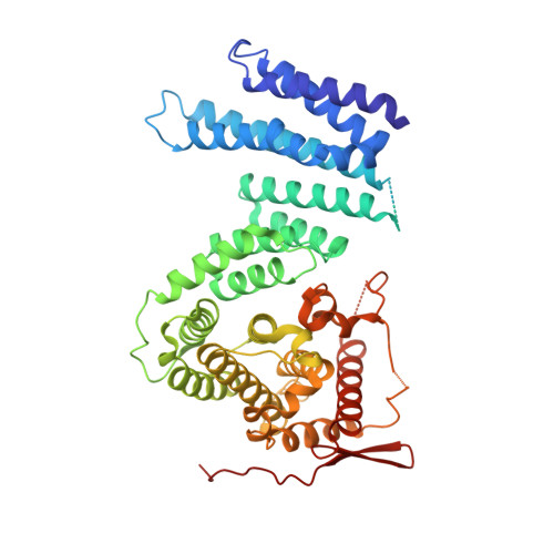
    1YA0

  • PubMed Abstract: 

    In metazoa, regulation of the phosphorylation state of UPF1 is crucial for nonsense-mediated mRNA decay (NMD), a process by which aberrant mRNAs containing nonsense mutations are degraded. UPF1 is targeted for dephosphorylation by three related proteins, SMG5, SMG6, and SMG7. We report here the crystal structure of the N-terminal domain of SMG7. The structure reveals that SMG7 contains a 14-3-3-like domain. Residues that bind phosphoserine-containing peptides in 14-3-3 are conserved at the equivalent positions in SMG7. Mutation of these residues impairs UPF1 binding to SMG7 in vitro and UPF1 recruitment to cytoplasmic mRNA decay foci in vivo, suggesting that SMG7 acts as an adaptor in targeting mRNAs associated with phosphorylated UPF1 for degradation. The 14-3-3 site of SMG7 is conserved in SMG5 and SMG6. These data also imply that the homologous human Est1 might have a 14-3-3 function at telomeres, and that phosphorylation events may be important for telomerase regulation.


  • Organizational Affiliation
    • European Molecular Biology Laboratory, Meyerhofstrasse 1, D-69117 Heidelberg, Germany.

Macromolecule Content 

  • Total Structure Weight: 114.63 kDa 
  • Atom Count: 7,854 
  • Modeled Residue Count: 922 
  • Deposited Residue Count: 994 
  • Unique protein chains: 1

Macromolecules

Find similar proteins by:|  3D Structure
Entity ID: 1
MoleculeChains  Sequence LengthOrganismDetailsImage
SMG-7 transcript variant 2
A, B
497Homo sapiensMutation(s): 0 
UniProt & NIH Common Fund Data Resources
Find proteins for Q92540 (Homo sapiens)
Explore Q92540 
Go to UniProtKB:  Q92540
PHAROS:  Q92540
GTEx:  ENSG00000116698 
Entity Groups
Sequence Clusters30% Identity50% Identity70% Identity90% Identity95% Identity100% Identity
UniProt GroupQ92540
Sequence Annotations
Expand
Reference Sequence

Experimental Data & Validation

Experimental Data

  • Method: X-RAY DIFFRACTION
  • Resolution: 2.55 Å
  • R-Value Free:  0.287 (Depositor), 0.290 (DCC) 
  • R-Value Work:  0.246 (Depositor), 0.249 (DCC) 
  • R-Value Observed: 0.250 (Depositor) 
Space Group: P 21 21 21
Unit Cell:
Length ( Å )Angle ( ˚ )
a = 64.018α = 90
b = 100.0497β = 90
c = 208.6214γ = 90
Software Package:
Software NamePurpose
MOSFLMdata reduction
SCALAdata scaling
SOLVEphasing
CNSrefinement
CCP4data scaling

Structure Validation

View Full Validation Report


Entry History 

Deposition Data

Revision History  (Full details and data files)

  • Version 1.0: 2005-03-15
    Type: Initial release
  • Version 1.1: 2008-04-30
    Changes: Version format compliance
  • Version 1.2: 2011-07-13
    Changes: Version format compliance
  • Version 1.3: 2024-02-14
    Changes: Data collection, Database references, Derived calculations