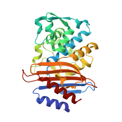
1YMX

X-ray crystallographic structure of CTX-M-9 beta-lactamase covalently linked to cefoxitin


Experimental Data Snapshot

  • Method: X-RAY DIFFRACTION
  • Resolution: 1.70 Å
  • R-Value Free: 0.197 
  • R-Value Work: 0.158 
  • R-Value Observed: 0.158 

Starting Model: experimental
View more details

wwPDB Validation   3D Report Full Report


Ligand Structure Quality Assessment 


This is version 2.2 of the entry. See complete history


Literature

Structure, Function, and Inhibition along the Reaction Coordinate of CTX-M beta-Lactamases.

Chen, Y.Shoichet, B.Bonnet, R.

(2005) J Am Chem Soc 127: 5423-5434

  • DOI: https://doi.org/10.1021/ja042850a
  • Primary Citation of Related Structures:  
    1YLY, 1YLZ, 1YM1, 1YMS, 1YMX

  • PubMed Abstract: 

    CTX-M enzymes are an emerging group of extended spectrum beta-lactamases (ESBLs) that hydrolyze not only the penicillins but also the first-, second-, and third-generation cephalosporins. Although they have become the most frequently observed ESBLs in certain areas, there are few effective inhibitors and relatively little is known about their detailed mechanism. Here we describe the X-ray crystal structures of CTX-M enzymes in complex with different transition-state analogues and beta-lactam inhibitors, representing the enzyme as it progresses from its acylation transition state to its acyl enzyme complex to the deacylation transition state. As the enzyme moves along this reaction coordinate, two key catalytic residues, Lys73 and Glu166, change conformations, tracking the state of the reaction. Unexpectedly, the acyl enzyme complex with the beta-lactam inhibitor cefoxitin still has the catalytic water bound; this water had been predicted to be displaced by the unusual 7alpha-methoxy of the inhibitor. Instead, the 7alpha-group appears to inhibit by preventing the formation of the deacylation transition state through steric hindrance. From an inhibitor design standpoint, we note that the best of the reversible inhibitors, a ceftazidime-like boronic acid compound, binds to CTX-M-16 with a K(i) value of 4 nM. When used together in cell culture, this inhibitor reversed cefotaxime resistance in CTX-M-producing bacteria. The structure of its complex with CTX-M enzyme and the structural view of the reaction coordinate described here provide templates for inhibitor design and intervention to combat this family of antibiotic resistance enzymes.


  • Organizational Affiliation

    Department of Pharmaceutical Chemistry, University of California, San Francisco, Genentech Hall, 600 16th Street, San Francisco, California 94143-2240, USA.


Macromolecules
Find similar proteins by:  (by identity cutoff)  |  3D Structure
Entity ID: 1
MoleculeChains Sequence LengthOrganismDetailsImage
beta-lactamase CTX-M-9
A, B
263Escherichia coliMutation(s): 0 
Gene Names: CTX-M
EC: 3.5.2.6
UniProt
Find proteins for Q9L5C8 (Escherichia coli)
Explore Q9L5C8 
Go to UniProtKB:  Q9L5C8
Entity Groups  
Sequence Clusters30% Identity50% Identity70% Identity90% Identity95% Identity100% Identity
UniProt GroupQ9L5C8
Sequence Annotations
Expand
  • Reference Sequence
Small Molecules
Ligands 1 Unique
IDChains Name / Formula / InChI Key2D Diagram3D Interactions
1QL
Query on 1QL

Download Ideal Coordinates CCD File 
C [auth A],
D [auth B]
(2R)-5-[(carbamoyloxy)methyl]-2-{(1S)-1-methoxy-2-oxo-1-[(thiophen-2-ylacetyl)amino]ethyl}-3,6-dihydro-2H-1,3-thiazine-4-carboxylic acid
C16 H19 N3 O7 S2
IYZWSAWEHPQLHS-ZBFHGGJFSA-N
Binding Affinity Annotations 
IDSourceBinding Affinity
CFX PDBBind:  1YMX IC50: 1.40e+4 (nM) from 1 assay(s)
Experimental Data & Validation

Experimental Data

  • Method: X-RAY DIFFRACTION
  • Resolution: 1.70 Å
  • R-Value Free: 0.197 
  • R-Value Work: 0.158 
  • R-Value Observed: 0.158 
  • Space Group: P 1 21 1
Unit Cell:
Length ( Å )Angle ( ˚ )
a = 45.209α = 90
b = 106.781β = 101.62
c = 47.779γ = 90
Software Package:
Software NamePurpose
CNSrefinement
DENZOdata reduction
SCALEPACKdata scaling
CNSphasing

Structure Validation

View Full Validation Report


Ligand Structure Quality Assessment 


Entry History 

Deposition Data

Revision History  (Full details and data files)

  • Version 1.0: 2005-04-26
    Type: Initial release
  • Version 1.1: 2008-04-30
    Changes: Version format compliance
  • Version 1.2: 2011-07-13
    Changes: Version format compliance
  • Version 2.0: 2018-09-12
    Changes: Advisory, Atomic model, Data collection, Derived calculations, Non-polymer description, Structure summary
  • Version 2.1: 2023-08-23
    Changes: Data collection, Database references, Derived calculations, Refinement description
  • Version 2.2: 2024-10-16
    Changes: Structure summary