1Z84

X-ray structure of galt-like protein from arabidopsis thaliana at5g18200


Experimental Data Snapshot

  • Method: X-RAY DIFFRACTION
  • Resolution: 1.83 Å
  • R-Value Free: 
    0.231 (Depositor), 0.230 (DCC) 
  • R-Value Work: 
    0.186 (Depositor), 0.190 (DCC) 
  • R-Value Observed: 
    0.188 (Depositor) 

Starting Model: experimental
View more details

wwPDB Validation   3D Report Full Report


Ligand Structure Quality Assessment 

Created with Raphaël 2.3.0Worse 01 BetterLigand structure goodness of fit to experimental dataBest fitted AMPClick on this verticalbar to view details

This is version 1.5 of the entry. See complete history

Re-refinement Note

A newer entry is available that reflects an alternative modeling of the original data: 2Q4H


Literature

Structure and Mechanism of an ADP-Glucose Phosphorylase from Arabidopsis thaliana

MCCOY, J.G.ARABSHAHI, A.BITTO, E.BINGMAN, C.A.RUZICKA, F.J.FREY, P.A.PHILLIPS JR., G.N.

(2006) Biochemistry 45: 3154-3162

  • DOI: https://doi.org/10.1021/bi052232m
  • Primary Citation of Related Structures:  
    1Z84, 1ZWJ

  • PubMed Abstract: 

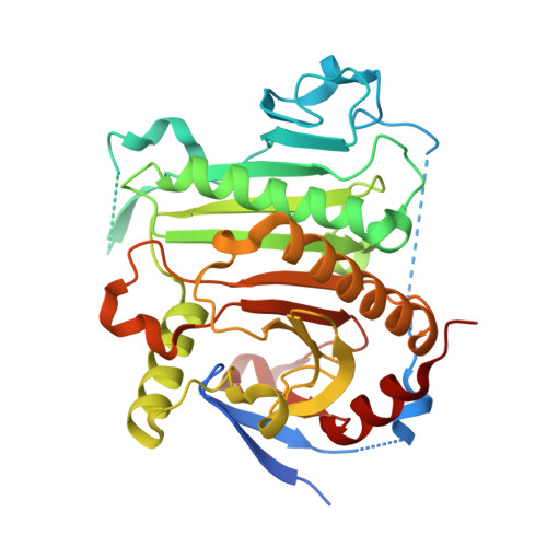
    The X-ray crystal structure of the At5g18200.1 protein has been determined to a nominal resolution of 2.30 A. The structure has a histidine triad (HIT)-like fold containing two distinct HIT-like motifs. The sequence of At5g18200.1 indicates a distant family relationship to the Escherichia coli galactose-1-P uridylyltransferase (GalT): the determined structure of the At5g18200.1 protein confirms this relationship. The At5g18200.1 protein does not demonstrate GalT activity but instead catalyzes adenylyl transfer in the reaction of ADP-glucose with various phosphates. The best acceptor among those evaluated is phosphate itself; thus, the At5g18200.1 enzyme appears to be an ADP-glucose phosphorylase. The enzyme catalyzes the exchange of (14)C between ADP-[(14)C]glucose and glucose-1-P in the absence of phosphate. The steady state kinetics of exchange follows the ping-pong bi-bi kinetic mechanism, with a k(cat) of 4.1 s(-)(1) and K(m) values of 1.4 and 83 microM for ADP-[(14)C]glucose and glucose-1-P, respectively, at pH 8.5 and 25 degrees C. The overall reaction of ADP-glucose with phosphate to produce ADP and glucose-1-P follows ping-pong bi-bi steady state kinetics, with a k(cat) of 2.7 s(-)(1) and K(m) values of 6.9 and 90 microM for ADP-glucose and phosphate, respectively, at pH 8.5 and 25 degrees C. The kinetics are consistent with a double-displacement mechanism that involves a covalent adenylyl-enzyme intermediate. The X-ray crystal structure of this intermediate was determined to 1.83 A resolution and shows the AMP group bonded to His(186). The value of K(eq) in the direction of ADP and glucose-1-P formation is 5.0 at pH 7.0 and 25 degrees C in the absence of a divalent metal ion, and it is 40 in the presence of 1 mM MgCl(2).


  • Organizational Affiliation

    Department of Biochemistry, University of Wisconsin, Madison, Wisconsin 53706, USA.


Macromolecules
Find similar proteins by:  (by identity cutoff)  |  3D Structure
Entity ID: 1
MoleculeChains Sequence LengthOrganismDetailsImage
galactose-1-phosphate uridyl transferase-like protein
A, B
351Arabidopsis thalianaMutation(s): 0 
Gene Names: AT5G18200
EC: 2.7.7
UniProt
Find proteins for Q9FK51 (Arabidopsis thaliana)
Explore Q9FK51 
Go to UniProtKB:  Q9FK51
Entity Groups  
Sequence Clusters30% Identity50% Identity70% Identity90% Identity95% Identity100% Identity
UniProt GroupQ9FK51
Sequence Annotations
Expand
  • Reference Sequence
Small Molecules
Ligands 3 Unique
IDChains Name / Formula / InChI Key2D Diagram3D Interactions
AMP
Query on AMP

Download Ideal Coordinates CCD File 
E [auth A],
K [auth B]
ADENOSINE MONOPHOSPHATE
C10 H14 N5 O7 P
UDMBCSSLTHHNCD-KQYNXXCUSA-N
ZN
Query on ZN

Download Ideal Coordinates CCD File 
C [auth A],
D [auth A],
I [auth B],
J [auth B]
ZINC ION
Zn
PTFCDOFLOPIGGS-UHFFFAOYSA-N
EDO
Query on EDO

Download Ideal Coordinates CCD File 
F [auth A]
G [auth A]
H [auth A]
L [auth B]
M [auth B]
1,2-ETHANEDIOL
C2 H6 O2
LYCAIKOWRPUZTN-UHFFFAOYSA-N
Experimental Data & Validation

Experimental Data

  • Method: X-RAY DIFFRACTION
  • Resolution: 1.83 Å
  • R-Value Free:  0.231 (Depositor), 0.230 (DCC) 
  • R-Value Work:  0.186 (Depositor), 0.190 (DCC) 
  • R-Value Observed: 0.188 (Depositor) 
Space Group: P 21 21 21
Unit Cell:
Length ( Å )Angle ( ˚ )
a = 62.037α = 90
b = 95.65β = 90
c = 110.918γ = 90
Software Package:
Software NamePurpose
DENZOdata reduction
SCALEPACKdata scaling
MOLREPphasing
REFMACrefinement

Structure Validation

View Full Validation Report


Ligand Structure Quality Assessment 

Created with Raphaël 2.3.0Worse 01 BetterLigand structure goodness of fit to experimental dataBest fitted AMPClick on this verticalbar to view details

Entry History 

Deposition Data

Revision History  (Full details and data files)

  • Version 1.0: 2005-04-19
    Type: Initial release
  • Version 1.1: 2008-04-30
    Changes: Version format compliance
  • Version 1.2: 2011-07-13
    Changes: Version format compliance
  • Version 1.3: 2017-10-11
    Changes: Refinement description
  • Version 1.4: 2023-08-23
    Changes: Data collection, Database references, Derived calculations, Refinement description
  • Version 1.5: 2024-10-16
    Changes: Structure summary