1ZR2 | pdb_00001zr2

Structure of a Synaptic gamma-delta Resolvase Tetramer Covalently Linked to two Cleaved DNAs


Experimental Data Snapshot

  • Method: X-RAY DIFFRACTION
  • Resolution: 3.90 Å
  • R-Value Free: 
    0.322 (Depositor), 0.295 (DCC) 
  • R-Value Work: 
    0.264 (Depositor), 0.249 (DCC) 
  • R-Value Observed: 
    0.270 (Depositor) 

wwPDB Validation 3D Report Full Report

Validation slider image for 1ZR2

This is version 1.5 of the entry. See complete history

Literature

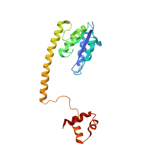
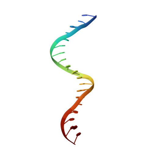
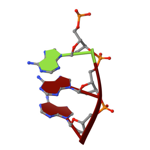
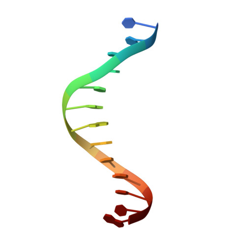
Structure of a synaptic gamma delta resolvase tetramer covalently linked to two cleaved DNAs.

Li, W.Kamtekar, S.Xiong, Y.Sarkis, G.J.Grindley, N.D.Steitz, T.A.

(2005) Science 309: 1210-1215

  • DOI: https://doi.org/10.1126/science.1112064
  • Primary Citation Related Structures: 
    1ZR2, 1ZR4

  • PubMed Abstract: 

    The structure of a synaptic intermediate of the site-specific recombinase gammadelta resolvase covalently linked through Ser10 to two cleaved duplex DNAs has been determined at 3.4 angstrom resolution. This resolvase, activated for recombination by mutations, forms a tetramer whose structure is substantially changed from that of a presynaptic complex between dimeric resolvase and the cleavage site DNA. Because the two cleaved DNA duplexes that are to be recombined lie on opposite sides of the core tetramer, large movements of both protein and DNA are required to achieve strand exchange. The two dimers linked to the DNAs that are to be recombined are held together by a flat interface. This may allow a 180 degrees rotation of one dimer relative to the other in order to reposition the DNA duplexes for strand exchange.


  • Organizational Affiliation
    • Department of Molecular Biophysics and Biochemistry, Yale University, New Haven, CT 06520, USA.

Macromolecule Content 

  • Total Structure Weight: 62.05 kDa 
  • Atom Count: 4,260 
  • Modeled Residue Count: 435 
  • Deposited Residue Count: 436 
  • Unique protein chains: 1
  • Unique nucleic acid chains: 3

Macromolecules


Find similar proteins by:|  3D Structure
Entity ID: 4
MoleculeChains  Sequence LengthOrganismDetailsImage
Transposon gamma-delta resolvaseG [auth A],
H [auth B]
183Escherichia coliMutation(s): 6 
Gene Names: tnpR
UniProt
Find proteins for P03012 (Escherichia coli (strain K12))
Explore P03012 
Go to UniProtKB:  P03012
Entity Groups
Sequence Clusters30% Identity50% Identity70% Identity90% Identity95% Identity100% Identity
UniProt GroupP03012
Sequence Annotations
Expand
Reference Sequence
Find similar nucleic acids by:  Sequence
Entity ID: 1
MoleculeChains LengthOrganismImage
TCAGTGTCCGATAATTTATA [auth X],
D [auth J]
19N/A
Sequence Annotations
Expand
Reference Sequence
Find similar nucleic acids by:  Sequence
Entity ID: 2
MoleculeChains LengthOrganismImage
AAAB [auth Z],
E [auth I]
3N/A
Sequence Annotations
Expand
Reference Sequence
Find similar nucleic acids by:  Sequence
Entity ID: 3
MoleculeChains LengthOrganismImage
TTATCGGACACTGC [auth Y],
F [auth K]
13N/A
Sequence Annotations
Expand
Reference Sequence

Experimental Data & Validation

Experimental Data

  • Method: X-RAY DIFFRACTION
  • Resolution: 3.90 Å
  • R-Value Free:  0.322 (Depositor), 0.295 (DCC) 
  • R-Value Work:  0.264 (Depositor), 0.249 (DCC) 
  • R-Value Observed: 0.270 (Depositor) 
Space Group: P 32 2 1
Unit Cell:
Length ( Å )Angle ( ˚ )
a = 125.05α = 90
b = 125.05β = 90
c = 127.39γ = 120
Software Package:
Software NamePurpose
REFMACrefinement
HKL-2000data reduction
SCALEPACKdata scaling
SOLVEphasing

Structure Validation

View Full Validation Report


Entry History 

Deposition Data

Revision History  (Full details and data files)

  • Version 1.0: 2005-08-30
    Type: Initial release
  • Version 1.1: 2008-04-30
    Changes: Version format compliance
  • Version 1.2: 2011-07-13
    Changes: Advisory, Version format compliance
  • Version 1.3: 2018-01-31
    Changes: Experimental preparation
  • Version 1.4: 2021-10-20
    Changes: Database references, Derived calculations
  • Version 1.5: 2024-10-16
    Changes: Data collection, Refinement description, Structure summary