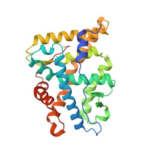
1A28

HORMONE-BOUND HUMAN PROGESTERONE RECEPTOR LIGAND-BINDING DOMAIN


Experimental Data Snapshot

  • Method: X-RAY DIFFRACTION
  • Resolution: 1.80 Å
  • R-Value Free: 0.227 
  • R-Value Work: 0.191 
  • R-Value Observed: 0.191 

wwPDB Validation   3D Report Full Report


Ligand Structure Quality Assessment 


This is version 1.3 of the entry. See complete history


Literature

Atomic structure of progesterone complexed with its receptor.

Williams, S.P.Sigler, P.B.

(1998) Nature 393: 392-396

  • DOI: https://doi.org/10.1038/30775
  • Primary Citation of Related Structures:  
    1A28

  • PubMed Abstract: 

    The physiological effects of progestins are mediated by the progesterone receptor, a member of the steroid/nuclear receptor superfamily. As progesterone is required for maintenance of pregnancy, its receptor has been a target for pharmaceuticals. Here we report the 1.8 A crystal structure of a progesterone-bound ligand-binding domain of the human progesterone receptor. The nature of this structure explains the receptor's selective affinity for progestins and establishes a common mode of recognition of 3-oxy steroids by the cognate receptors. Although the overall fold of the progesterone receptor is similar to that found in related receptors, the progesterone receptor has a quite different mode of dimerization. A hormone-induced stabilization of the carboxy-terminal secondary structure of the ligand-binding domain of the progesterone receptor accounts for the stereochemistry of this distinctive dimer, explains the receptor's characteristic pattern of ligand-dependent protease resistance and its loss of repression, and indicates how the anti-progestin RU486 might work in birth control. The structure also indicates that the analogous 3-keto-steroid receptors may have a similar mechanism of action.


  • Organizational Affiliation

    Department of Molecular Biophysics and Biochemistry, and the Howard Hughes Medical Institute, Yale University, New Haven, Connecticut 06510, USA.


Macromolecules
Find similar proteins by:  (by identity cutoff)  |  3D Structure
Entity ID: 1
MoleculeChains Sequence LengthOrganismDetailsImage
PROGESTERONE RECEPTOR
A, B
256Homo sapiensMutation(s): 0 
Gene Names: PGR
UniProt & NIH Common Fund Data Resources
Find proteins for P06401 (Homo sapiens)
Explore P06401 
Go to UniProtKB:  P06401
PHAROS:  P06401
GTEx:  ENSG00000082175 
Entity Groups  
Sequence Clusters30% Identity50% Identity70% Identity90% Identity95% Identity100% Identity
UniProt GroupP06401
Sequence Annotations
Expand
  • Reference Sequence
Small Molecules
Binding Affinity Annotations 
IDSourceBinding Affinity
STR BindingDB:  1A28 Ki: min: 3.4, max: 10.4 (nM) from 5 assay(s)
IC50: min: 0.2, max: 17 (nM) from 6 assay(s)
EC50: min: 0.1, max: 50 (nM) from 12 assay(s)
PDBBind:  1A28 Ki: 5.1 (nM) from 1 assay(s)
Experimental Data & Validation

Experimental Data

  • Method: X-RAY DIFFRACTION
  • Resolution: 1.80 Å
  • R-Value Free: 0.227 
  • R-Value Work: 0.191 
  • R-Value Observed: 0.191 
  • Space Group: P 1 21 1
Unit Cell:
Length ( Å )Angle ( ˚ )
a = 58.123α = 90
b = 64.444β = 95.74
c = 69.954γ = 90
Software Package:
Software NamePurpose
DPHASEmodel building
CNSrefinement
DENZOdata reduction
SCALEPACKdata scaling
DPHASEphasing

Structure Validation

View Full Validation Report


Ligand Structure Quality Assessment 


Entry History 

Deposition Data

Revision History  (Full details and data files)

  • Version 1.0: 1998-07-15
    Type: Initial release
  • Version 1.1: 2008-03-24
    Changes: Version format compliance
  • Version 1.2: 2011-07-13
    Changes: Version format compliance
  • Version 1.3: 2024-02-07
    Changes: Data collection, Database references, Derived calculations, Other