1F6B

CRYSTAL STRUCTURE OF SAR1-GDP COMPLEX


Experimental Data Snapshot

  • Method: X-RAY DIFFRACTION
  • Resolution: 1.70 Å
  • R-Value Free: 0.240 
  • R-Value Work: 0.220 
  • R-Value Observed: 0.220 

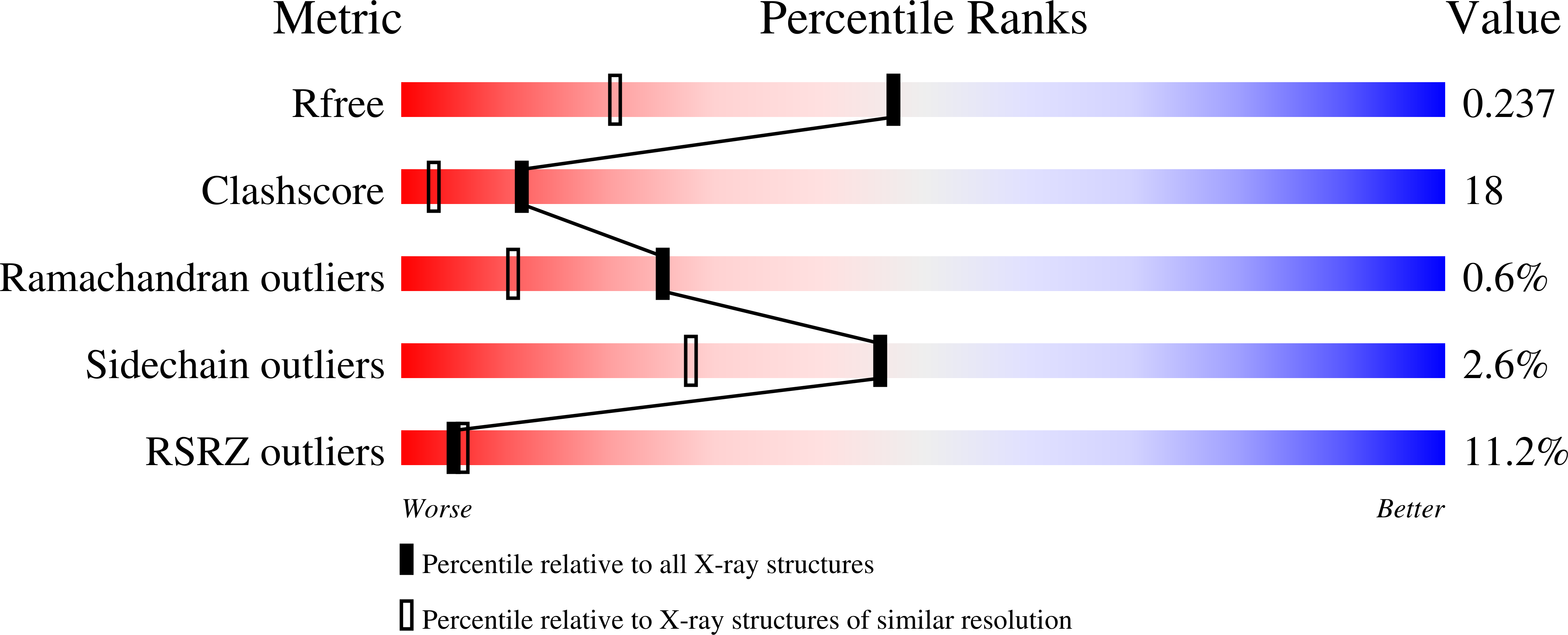
wwPDB Validation   3D Report Full Report


Ligand Structure Quality Assessment 


This is version 1.3 of the entry. See complete history


Literature

Crystal structure of Sar1-GDP at 1.7 A resolution and the role of the NH2 terminus in ER export.

Huang, M.Weissman, J.T.Beraud-Dufour, S.Luan, P.Wang, C.Chen, W.Aridor, M.Wilson, I.A.Balch, W.E.

(2001) J Cell Biol 155: 937-948

  • DOI: https://doi.org/10.1083/jcb.200106039
  • Primary Citation of Related Structures:  
    1F6B

  • PubMed Abstract: 

    The Sar1 GTPase is an essential component of COPII vesicle coats involved in export of cargo from the ER. We report the 1.7-A structure of Sar1 and find that consistent with the sequence divergence of Sar1 from Arf family GTPases, Sar1 is structurally distinct. In particular, we show that the Sar1 NH2 terminus contains two regions: an NH2-terminal extension containing an evolutionary conserved hydrophobic motif that facilitates membrane recruitment and activation by the mammalian Sec12 guanine nucleotide exchange factor, and an alpha1' amphipathic helix that contributes to interaction with the Sec23/24 complex that is responsible for cargo selection during ER export. We propose that the hydrophobic Sar1 NH2-terminal activation/recruitment motif, in conjunction with the alpha1' helix, mediates the initial steps in COPII coat assembly for export from the ER.


  • Organizational Affiliation

    Department of Cell Biology, The Scripps Research Institute, La Jolla, CA 92130, USA.


Macromolecules
Find similar proteins by:  (by identity cutoff)  |  3D Structure
Entity ID: 1
MoleculeChains Sequence LengthOrganismDetailsImage
SAR1
A, B
198Cricetulus griseusMutation(s): 0 
EC: 3.6.5.2
UniProt
Find proteins for Q9QVY3 (Cricetulus griseus)
Explore Q9QVY3 
Go to UniProtKB:  Q9QVY3
Entity Groups  
Sequence Clusters30% Identity50% Identity70% Identity90% Identity95% Identity100% Identity
UniProt GroupQ9QVY3
Sequence Annotations
Expand
  • Reference Sequence
Small Molecules
Ligands 3 Unique
IDChains Name / Formula / InChI Key2D Diagram3D Interactions
GDP
Query on GDP

Download Ideal Coordinates CCD File 
G [auth A],
K [auth B]
GUANOSINE-5'-DIPHOSPHATE
C10 H15 N5 O11 P2
QGWNDRXFNXRZMB-UUOKFMHZSA-N
SO4
Query on SO4

Download Ideal Coordinates CCD File 
D [auth A],
E [auth A],
F [auth A],
I [auth B],
J [auth B]
SULFATE ION
O4 S
QAOWNCQODCNURD-UHFFFAOYSA-L
MG
Query on MG

Download Ideal Coordinates CCD File 
C [auth A],
H [auth B]
MAGNESIUM ION
Mg
JLVVSXFLKOJNIY-UHFFFAOYSA-N
Experimental Data & Validation

Experimental Data

  • Method: X-RAY DIFFRACTION
  • Resolution: 1.70 Å
  • R-Value Free: 0.240 
  • R-Value Work: 0.220 
  • R-Value Observed: 0.220 
  • Space Group: P 1 21 1
Unit Cell:
Length ( Å )Angle ( ˚ )
a = 53.368α = 90
b = 61.689β = 107.49
c = 71.142γ = 90
Software Package:
Software NamePurpose
PHASESphasing
CNSrefinement
DENZOdata reduction
SCALEPACKdata scaling

Structure Validation

View Full Validation Report


Ligand Structure Quality Assessment 


Entry History 

Deposition Data

Revision History  (Full details and data files)

  • Version 1.0: 2002-01-09
    Type: Initial release
  • Version 1.1: 2008-04-27
    Changes: Version format compliance
  • Version 1.2: 2011-07-13
    Changes: Version format compliance
  • Version 1.3: 2024-02-07
    Changes: Data collection, Database references, Derived calculations