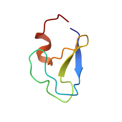
1PTQ

PROTEIN KINASE C DELTA CYS2 DOMAIN


Experimental Data Snapshot

  • Method: X-RAY DIFFRACTION
  • Resolution: 1.95 Å
  • R-Value Work: 0.196 
  • R-Value Observed: 0.196 

wwPDB Validation   3D Report Full Report


This is version 1.3 of the entry. See complete history


Literature

Crystal structure of the cys2 activator-binding domain of protein kinase C delta in complex with phorbol ester.

Zhang, G.Kazanietz, M.G.Blumberg, P.M.Hurley, J.H.

(1995) Cell 81: 917-924

  • DOI: https://doi.org/10.1016/0092-8674(95)90011-x
  • Primary Citation of Related Structures:  
    1PTQ, 1PTR

  • PubMed Abstract: 

    Protein kinase Cs (PKCs) are a ubiquitous family of regulatory enzymes that associate with membranes and are activated by diacylglycerol or tumor-promoting agonists such as phorbol esters. The structure of the second activator-binding domain of PKC delta has been determined in complex with phorbol 13-acetate, which binds in a groove between two pulled-apart beta strands at the tip of the domain. The C3, C4, and C20 phorbol oxygens form hydrogen bonds with main-chain groups whose orientation is controlled by a set of highly conserved residues. Phorbol binding caps the groove and forms a contiguous hydrophobic surface covering one-third of the domain, explaining how the activator promotes insertion of PKC into membranes.


  • Organizational Affiliation

    Laboratory of Molecular Biology, National Institute of Diabetes and Digestive and Kidney Diseases, National Institutes of Health, Bethesda, Maryland 20892-0580, USA.


Macromolecules
Find similar proteins by:  (by identity cutoff)  |  3D Structure
Entity ID: 1
MoleculeChains Sequence LengthOrganismDetailsImage
PROTEIN KINASE C DELTA TYPE50Mus musculusMutation(s): 0 
EC: 2.7.1 (PDB Primary Data), 2.7.11.13 (UniProt), 2.7.10.2 (UniProt)
UniProt
Find proteins for P28867 (Mus musculus)
Explore P28867 
Go to UniProtKB:  P28867
Entity Groups  
Sequence Clusters30% Identity50% Identity70% Identity90% Identity95% Identity100% Identity
UniProt GroupP28867
Sequence Annotations
Expand
  • Reference Sequence
Experimental Data & Validation

Experimental Data

  • Method: X-RAY DIFFRACTION
  • Resolution: 1.95 Å
  • R-Value Work: 0.196 
  • R-Value Observed: 0.196 
  • Space Group: C 2 2 21
Unit Cell:
Length ( Å )Angle ( ˚ )
a = 32.4α = 90
b = 63.5β = 90
c = 65.5γ = 90
Software Package:
Software NamePurpose
X-PLORmodel building
X-PLORrefinement
DENZOdata reduction
X-PLORphasing

Structure Validation

View Full Validation Report


Entry History 

Deposition Data

Revision History  (Full details and data files)

  • Version 1.0: 1995-07-31
    Type: Initial release
  • Version 1.1: 2008-03-24
    Changes: Version format compliance
  • Version 1.2: 2011-07-13
    Changes: Version format compliance
  • Version 1.3: 2024-02-14
    Changes: Data collection, Database references, Derived calculations, Other