1R20

Crystal structure of the ligand-binding domains of the heterodimer EcR/USP bound to the synthetic agonist BYI06830


Experimental Data Snapshot

  • Method: X-RAY DIFFRACTION
  • Resolution: 3.00 Å
  • R-Value Free: 
    0.289 (Depositor), 0.280 (DCC) 
  • R-Value Work: 
    0.244 (Depositor), 0.230 (DCC) 
  • R-Value Observed: 
    0.244 (Depositor) 

wwPDB Validation   3D Report Full Report


Ligand Structure Quality Assessment 

Created with Raphaël 2.3.0Worse 01 BetterLigand structure goodness of fit to experimental dataBest fitted EPHClick on this verticalbar to view detailsBest fitted HWGClick on this verticalbar to view details

This is version 1.3 of the entry. See complete history


Literature

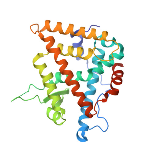
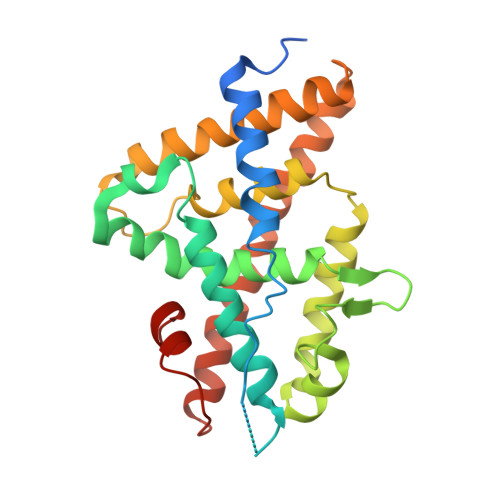
Structural adaptability in the ligand-binding pocket of the ecdysone hormone receptor.

Billas, I.M.L.Iwema, T.Garnier, J.M.Mitschler, A.Rochel, N.Moras, D.

(2003) Nature 426: 91-96

  • DOI: https://doi.org/10.1038/nature02112
  • Primary Citation of Related Structures:  
    1R1K, 1R20

  • PubMed Abstract: 

    The ecdysteroid hormones coordinate the major stages of insect development, notably moulting and metamorphosis, by binding to the ecdysone receptor (EcR); a ligand-inducible nuclear transcription factor. To bind either ligand or DNA, EcR must form a heterodimer with ultraspiracle (USP), the homologue of retinoid-X receptor. Here we report the crystal structures of the ligand-binding domains of the moth Heliothis virescens EcR-USP heterodimer in complex with the ecdysteroid ponasterone A and with a non-steroidal, lepidopteran-specific agonist BYI06830 used in agrochemical pest control. The two structures of EcR-USP emphasize the universality of heterodimerization as a general mechanism common to both vertebrates and invertebrates. Comparison of the EcR structures in complex with steroidal and non-steroidal ligands reveals radically different and only partially overlapping ligand-binding pockets that could not be predicted by molecular modelling and docking studies. These findings offer new perspectives for the design of insect-specific, environmentally safe insecticides. The concept of a ligand-dependent binding pocket in EcR provides an insight into the moulding of nuclear receptors to their ligand, and has potential applications for human nuclear receptors.


  • Organizational Affiliation

    Département de Biologie et de Génomique Structurales, IGBMC, CNRS/INSERM/Université Louis Pasteur, Parc d'Innovation BP10142, 67404 Illkirch cedex, France.


Macromolecules
Find similar proteins by:  (by identity cutoff)  |  3D Structure
Entity ID: 1
MoleculeChains Sequence LengthOrganismDetailsImage
ULTRASPIRACLE PROTEIN263Heliothis virescensMutation(s): 0 
UniProt
Find proteins for Q7SIF6 (Heliothis virescens)
Explore Q7SIF6 
Go to UniProtKB:  Q7SIF6
Entity Groups  
Sequence Clusters30% Identity50% Identity70% Identity90% Identity95% Identity100% Identity
UniProt GroupQ7SIF6
Sequence Annotations
Expand
  • Reference Sequence
Find similar proteins by:  (by identity cutoff)  |  3D Structure
Entity ID: 2
MoleculeChains Sequence LengthOrganismDetailsImage
ECDYSONE RECEPTORB [auth D]265Heliothis virescensMutation(s): 0 
UniProt
Find proteins for O18473 (Heliothis virescens)
Explore O18473 
Go to UniProtKB:  O18473
Entity Groups  
Sequence Clusters30% Identity50% Identity70% Identity90% Identity95% Identity100% Identity
UniProt GroupO18473
Sequence Annotations
Expand
  • Reference Sequence
Small Molecules
Ligands 2 Unique
IDChains Name / Formula / InChI Key2D Diagram3D Interactions
EPH
Query on EPH

Download Ideal Coordinates CCD File 
C [auth A]L-ALPHA-PHOSPHATIDYL-BETA-OLEOYL-GAMMA-PALMITOYL-PHOSPHATIDYLETHANOLAMINE
C39 H68 N O8 P
MABRTXOVHMDVAT-AAEGOEIASA-N
HWG
Query on HWG

Download Ideal Coordinates CCD File 
D
N-(TERT-BUTYL)-3,5-DIMETHYL-N'-[(5-METHYL-2,3-DIHYDRO-1,4-BENZODIOXIN-6-YL)CARBONYL]BENZOHYDRAZIDE
C23 H28 N2 O4
JGJACZABNQGPMT-UHFFFAOYSA-N
Experimental Data & Validation

Experimental Data

  • Method: X-RAY DIFFRACTION
  • Resolution: 3.00 Å
  • R-Value Free:  0.289 (Depositor), 0.280 (DCC) 
  • R-Value Work:  0.244 (Depositor), 0.230 (DCC) 
  • R-Value Observed: 0.244 (Depositor) 
Space Group: P 3 2 1
Unit Cell:
Length ( Å )Angle ( ˚ )
a = 149.429α = 90
b = 149.429β = 90
c = 61.058γ = 120
Software Package:
Software NamePurpose
HKL-2000data collection
SCALEPACKdata scaling
EPMRphasing
CNSrefinement
HKL-2000data reduction

Structure Validation

View Full Validation Report


Ligand Structure Quality Assessment 

Created with Raphaël 2.3.0Worse 01 BetterLigand structure goodness of fit to experimental dataBest fitted EPHClick on this verticalbar to view detailsBest fitted HWGClick on this verticalbar to view details

Entry History 

Deposition Data

Revision History  (Full details and data files)

  • Version 1.0: 2003-11-18
    Type: Initial release
  • Version 1.1: 2008-04-29
    Changes: Version format compliance
  • Version 1.2: 2011-07-13
    Changes: Version format compliance
  • Version 1.3: 2024-02-14
    Changes: Data collection, Database references, Derived calculations, Structure summary