1YSL | pdb_00001ysl

Crystal structure of HMG-CoA synthase from Enterococcus faecalis with AcetoAcetyl-CoA ligand.


Experimental Data Snapshot

  • Method: X-RAY DIFFRACTION
  • Resolution: 1.90 Å
  • R-Value Free: 
    0.201 (Depositor), 0.208 (DCC) 
  • R-Value Work: 
    0.179 (Depositor), 0.187 (DCC) 
  • R-Value Observed: 
    0.180 (Depositor) 

Starting Model: experimental
View more details

wwPDB Validation 3D Report Full Report

Validation slider image for 1YSL

Ligand Structure Quality Assessment 


This is version 1.5 of the entry. See complete history

Literature

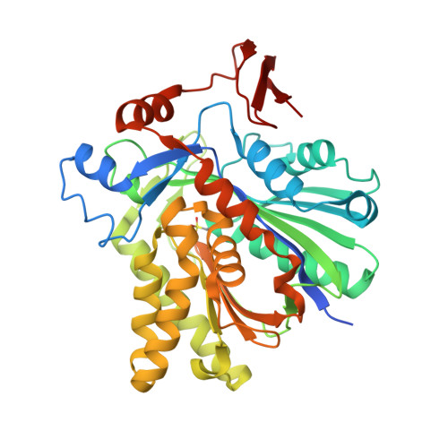
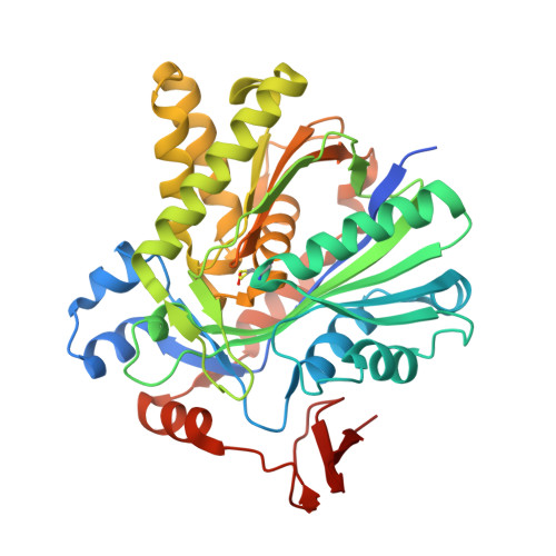
X-ray Crystal Structures of HMG-CoA Synthase from Enterococcus faecalis and a Complex with Its Second Substrate/Inhibitor Acetoacetyl-CoA.

Steussy, C.N.Vartia, A.A.Burgner II, J.W.Sutherlin, A.Rodwell, V.W.Stauffacher, C.V.

(2005) Biochemistry 44: 14256-14267

  • DOI: https://doi.org/10.1021/bi051487x
  • Primary Citation Related Structures: 
    1X9E, 1YSL

  • PubMed Abstract: 

    Biosynthesis of the isoprenoid precursor, isopentenyl diphosphate, is a critical function in all independently living organisms. There are two major pathways for this synthesis, the non-mevalonate pathway found in most eubacteria and the mevalonate pathway found in animal cells and a number of pathogenic bacteria. An early step in this pathway is the condensation of acetyl-CoA and acetoacetyl-CoA into HMG-CoA, catalyzed by the enzyme HMG-CoA synthase. To explore the possibility of a small molecule inhibitor of the enzyme functioning as a non-cell wall antibiotic, the structure of HMG-CoA synthase from Enterococcus faecalis (MVAS) was determined by selenomethionine MAD phasing to 2.4 A and the enzyme complexed with its second substrate, acetoacetyl-CoA, to 1.9 A. These structures show that HMG-CoA synthase from Enterococcus is a member of the family of thiolase fold enzymes and, while similar to the recently published HMG-CoA synthase structures from Staphylococcus aureus, exhibit significant differences in the structure of the C-terminal domain. The acetoacetyl-CoA binary structure demonstrates reduced coenzyme A and acetoacetate covalently bound to the active site cysteine through a thioester bond. This is consistent with the kinetics of the reaction that have shown acetoacetyl-CoA to be a potent inhibitor of the overall reaction, and provides a starting point in the search for a small molecule inhibitor.


  • Organizational Affiliation
    • Department of Biological Sciences, Purdue University, West Lafayette, Indiana 47907, USA.

Macromolecule Content 

  • Total Structure Weight: 89.83 kDa 
  • Atom Count: 6,670 
  • Modeled Residue Count: 767 
  • Deposited Residue Count: 805 
  • Unique protein chains: 2

Macromolecules

Find similar proteins by:|  3D Structure
Entity ID: 1
MoleculeChains  Sequence LengthOrganismDetailsImage
HMG-CoA synthase402Enterococcus faecalisMutation(s): 0 
EC: 4.1.3.5 (PDB Primary Data), 2.3.3.10 (UniProt)
UniProt
Find proteins for Q9FD71 (Enterococcus faecalis)
Explore Q9FD71 
Go to UniProtKB:  Q9FD71
Entity Groups
Sequence Clusters30% Identity50% Identity70% Identity90% Identity95% Identity100% Identity
UniProt GroupQ9FD71
Sequence Annotations
Expand
Reference Sequence
Find similar proteins by:|  3D Structure
Entity ID: 2
MoleculeChains  Sequence LengthOrganismDetailsImage
HMG-CoA synthase402Enterococcus faecalisMutation(s): 1 
EC: 4.1.3.5 (PDB Primary Data), 2.3.3.10 (UniProt)
UniProt
Find proteins for Q9FD71 (Enterococcus faecalis)
Explore Q9FD71 
Go to UniProtKB:  Q9FD71
Entity Groups
Sequence Clusters30% Identity50% Identity70% Identity90% Identity95% Identity100% Identity
UniProt GroupQ9FD71
Sequence Annotations
Expand
Reference Sequence

Small Molecules

Ligands 4 Unique
IDChains Name / Formula / InChI Key2D Diagram3D Interactions
COA

Query on COA


Download:Ideal Coordinates CCD File
F [auth B]COENZYME A
C21 H36 N7 O16 P3 S
RGJOEKWQDUBAIZ-IBOSZNHHSA-N
AAE

Query on AAE


Download:Ideal Coordinates CCD File
G [auth B]ACETOACETIC ACID
C4 H6 O3
WDJHALXBUFZDSR-UHFFFAOYSA-N
SO4

Query on SO4


Download:Ideal Coordinates CCD File
C [auth A],
E [auth B]
SULFATE ION
O4 S
QAOWNCQODCNURD-UHFFFAOYSA-L
GOL

Query on GOL


Download:Ideal Coordinates CCD File
D [auth A],
H [auth B]
GLYCEROL
C3 H8 O3
PEDCQBHIVMGVHV-UHFFFAOYSA-N
Modified Residues  1 Unique
IDChains TypeFormula2D DiagramParent
CSD
Query on CSD
A
L-PEPTIDE LINKINGC3 H7 N O4 SCYS

Experimental Data & Validation

Experimental Data

  • Method: X-RAY DIFFRACTION
  • Resolution: 1.90 Å
  • R-Value Free:  0.201 (Depositor), 0.208 (DCC) 
  • R-Value Work:  0.179 (Depositor), 0.187 (DCC) 
  • R-Value Observed: 0.180 (Depositor) 
Space Group: I 2 2 2
Unit Cell:
Length ( Å )Angle ( ˚ )
a = 105.3α = 90
b = 109.013β = 90
c = 140.799γ = 90
Software Package:
Software NamePurpose
HKL-2000data collection
SCALEPACKdata scaling
CNSrefinement
HKL-2000data reduction
CNSphasing

Structure Validation

View Full Validation Report


Ligand Structure Quality Assessment 


Entry History 

Deposition Data

Revision History  (Full details and data files)

  • Version 1.0: 2005-11-08
    Type: Initial release
  • Version 1.1: 2008-04-30
    Changes: Version format compliance
  • Version 1.2: 2011-07-13
    Changes: Non-polymer description, Version format compliance
  • Version 1.3: 2011-11-16
    Changes: Atomic model
  • Version 1.4: 2023-08-23
    Changes: Data collection, Database references, Derived calculations, Refinement description, Structure summary
  • Version 1.5: 2024-12-25
    Changes: Advisory, Derived calculations, Structure summary