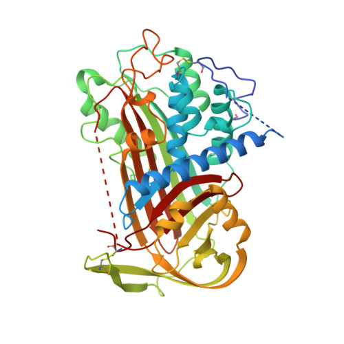
2ANT

THE 2.6 A STRUCTURE OF ANTITHROMBIN INDICATES A CONFORMATIONAL CHANGE AT THE HEPARIN BINDING SITE


Experimental Data Snapshot

  • Method: X-RAY DIFFRACTION
  • Resolution: 2.60 Å
  • R-Value Free: 0.290 
  • R-Value Work: 0.217 

Starting Model: experimental
View more details

wwPDB Validation   3D Report Full Report


Ligand Structure Quality Assessment 


This is version 1.4 of the entry. See complete history


Literature

The 2.6 A structure of antithrombin indicates a conformational change at the heparin binding site.

Skinner, R.Abrahams, J.P.Whisstock, J.C.Lesk, A.M.Carrell, R.W.Wardell, M.R.

(1997) J Mol Biol 266: 601-609

  • DOI: https://doi.org/10.1006/jmbi.1996.0798
  • Primary Citation of Related Structures:  
    2ANT

  • PubMed Abstract: 

    The crystal structure of a dimeric form of intact antithrombin has been solved to 2.6 A, representing the highest-resolution structure of an active, inhibitory serpin to date. The crystals were grown under microgravity conditions on Space Shuttle mission STS-67. The overall confidence in the structure, determined earlier from lower resolution data, is increased and new insights into the structure-function relationship are gained. Clear and continuous electron density is present for the reactive centre loop region P12 to P14 inserting into the top of the A-beta-sheet. Areas of the extended amino terminus, unique to antithrombin and important in the binding of the glycosaminoglycan heparin, can now be traced further than in the earlier structures. As in the earlier studies, the crystals contain one active and one latent molecule per asymmetric unit. Better definition of the electron density surrounding the D-helix and of the residues implicated in the binding of the heparin pentasaccharide (Arg47, Lys114, Lys125, Arg129) provides an insight into the change of affinity of binding that accompanies the change in conformation. In particular, the observed hydrogen bonding of these residues to the body of the molecule in the latent form explains the mechanism for the release of newly formed antithrombin-protease complexes into the circulation for catabolic removal.


  • Organizational Affiliation

    Department of Haematology, University of Cambridge, MRC Centre, UK.


Macromolecules
Find similar proteins by:  (by identity cutoff)  |  3D Structure
Entity ID: 1
MoleculeChains Sequence LengthOrganismDetailsImage
ANTITHROMBINA [auth L],
B [auth I]
432Homo sapiensMutation(s): 0 
UniProt & NIH Common Fund Data Resources
Find proteins for P01008 (Homo sapiens)
Explore P01008 
Go to UniProtKB:  P01008
PHAROS:  P01008
GTEx:  ENSG00000117601 
Entity Groups  
Sequence Clusters30% Identity50% Identity70% Identity90% Identity95% Identity100% Identity
UniProt GroupP01008
Sequence Annotations
Expand
  • Reference Sequence
Experimental Data & Validation

Experimental Data

  • Method: X-RAY DIFFRACTION
  • Resolution: 2.60 Å
  • R-Value Free: 0.290 
  • R-Value Work: 0.217 
  • Space Group: P 1 21 1
Unit Cell:
Length ( Å )Angle ( ˚ )
a = 61.41α = 90
b = 98.31β = 103.32
c = 90.41γ = 90
Software Package:
Software NamePurpose
MOSFLMdata reduction
SCALAdata scaling
AMoREphasing
TNTrefinement
CCP4data scaling

Structure Validation

View Full Validation Report


Ligand Structure Quality Assessment 


Entry History 

Deposition Data

Revision History  (Full details and data files)

  • Version 1.0: 1997-06-16
    Type: Initial release
  • Version 1.1: 2008-03-24
    Changes: Version format compliance
  • Version 1.2: 2011-07-13
    Changes: Version format compliance
  • Version 1.3: 2020-07-29
    Type: Remediation
    Reason: Carbohydrate remediation
    Changes: Advisory, Data collection, Derived calculations, Structure summary
  • Version 1.4: 2023-08-09
    Changes: Advisory, Database references, Refinement description, Structure summary