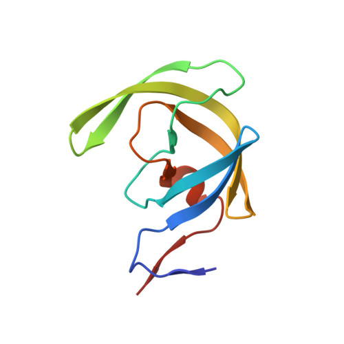
Molecular basis for substrate recognition and drug resistance from 1.1 to 1.6 angstroms resolution crystal structures of HIV-1 protease mutants with substrate analogs.
Tie, Y., Boross, P.I., Wang, Y.F., Gaddis, L., Liu, F., Chen, X., Tozser, J., Harrison, R.W., Weber, I.T.(2005) FEBS J 272: 5265-5277
- PubMed: 16218957
- DOI: https://doi.org/10.1111/j.1742-4658.2005.04923.x
- Primary Citation of Related Structures:
2AOC, 2AOD, 2AOE, 2AOF, 2AOG, 2AOH, 2AOI, 2AOJ - PubMed Abstract:
HIV-1 protease (PR) and two drug-resistant variants--PR with the V82A mutation (PR(V82A)) and PR with the I84V mutation (PR(I84V))--were studied using reduced peptide analogs of five natural cleavage sites (CA-p2, p2-NC, p6pol-PR, p1-p6 and NC-p1) to understand the structural and kinetic changes. The common drug-resistant mutations V82A and I84V alter residues forming the substrate-binding site. Eight crystal structures were refined at resolutions of 1.10-1.60 A. Differences in the PR-analog interactions depended on the peptide sequence and were consistent with the relative inhibition. Analog p6(pol)-PR formed more hydrogen bonds of P2 Asn with PR and fewer van der Waals contacts at P1' Pro compared with those formed by CA-p2 or p2-NC in PR complexes. The P3 Gly in p1-p6 provided fewer van der Waals contacts and hydrogen bonds at P2-P3 and more water-mediated interactions. PR(I84V) showed reduced van der Waals interactions with inhibitor compared with PR, which was consistent with kinetic data. The structures suggest that the binding affinity for mutants is modulated by the conformational flexibility of the substrate analogs. The complexes of PR(V82A) showed smaller shifts of the main chain atoms of Ala82 relative to PR, but more movement of the peptide analog, compared to complexes with clinical inhibitors. PR(V82A) was able to compensate for the loss of interaction with inhibitor caused by mutation, in agreement with kinetic data, but substrate analogs have more flexibility than the drugs to accommodate the structural changes caused by mutation. Hence, these structures help to explain how HIV can develop drug resistance while retaining the ability of PR to hydrolyze natural substrates.
- Department of Chemistry, Molecular Basis of Disease, Georgia State University, Atlanta, GA 30302-4010, USA.
Organizational Affiliation:


