2ATC | pdb_00002atc

CRYSTAL AND MOLECULAR STRUCTURES OF NATIVE AND CTP-LIGANDED ASPARTATE CARBAMOYLTRANSFERASE FROM ESCHERICHIA COLI


Experimental Data Snapshot

  • Method: X-RAY DIFFRACTION
  • Resolution: 3.00 Å
  • R-Value Work: 
    0.270 (Depositor) 

wwPDB Validation 3D Report Full Report

Validation slider image for 2ATC

This is version 1.4 of the entry. See complete history

Literature

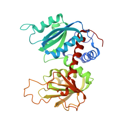
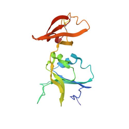
Crystal and molecular structures of native and CTP-liganded aspartate carbamoyltransferase from Escherichia coli.

Honzatko, R.B.Crawford, J.L.Monaco, H.L.Ladner, J.E.Ewards, B.F.Evans, D.R.Warren, S.G.Wiley, D.C.Ladner, R.C.Lipscomb, W.N.

(1982) J Mol Biology 160: 219-263

Macromolecule Content 

  • Total Structure Weight: 50.63 kDa 
  • Atom Count: 3,511 
  • Modeled Residue Count: 457 
  • Deposited Residue Count: 457 
  • Unique protein chains: 2

Macromolecules

Find similar proteins by:|  3D Structure
Entity ID: 1
MoleculeChains  Sequence LengthOrganismDetailsImage
ASPARTATE CARBAMOYLTRANSFERASE, CATALYTIC CHAIN305Escherichia coliMutation(s): 0 
EC: 2.1.3.2
UniProt
Find proteins for P0A786 (Escherichia coli (strain K12))
Explore P0A786 
Go to UniProtKB:  P0A786
Entity Groups
Sequence Clusters30% Identity50% Identity70% Identity90% Identity95% Identity100% Identity
UniProt GroupP0A786
Sequence Annotations
Expand
Reference Sequence
Find similar proteins by:|  3D Structure
Entity ID: 2
MoleculeChains  Sequence LengthOrganismDetailsImage
ASPARTATE CARBAMOYLTRANSFERASE, REGULATORY CHAIN152Escherichia coliMutation(s): 0 
UniProt
Find proteins for P0A7F3 (Escherichia coli (strain K12))
Explore P0A7F3 
Go to UniProtKB:  P0A7F3
Entity Groups
Sequence Clusters30% Identity50% Identity70% Identity90% Identity95% Identity100% Identity
UniProt GroupP0A7F3
Sequence Annotations
Expand
Reference Sequence

Small Molecules

Ligands 1 Unique
IDChains Name / Formula / InChI Key2D Diagram3D Interactions
ZN

Query on ZN


Download:Ideal Coordinates CCD File
C [auth B]ZINC ION
Zn
PTFCDOFLOPIGGS-UHFFFAOYSA-N

Experimental Data & Validation

Experimental Data

  • Method: X-RAY DIFFRACTION
  • Resolution: 3.00 Å
  • R-Value Work:  0.270 (Depositor) 
Space Group: H 3 2
Unit Cell:
Length ( Å )Angle ( ˚ )
a = 131.7α = 90
b = 131.7β = 90
c = 199.2γ = 120
Software Package:
Software NamePurpose
PROLSQrefinement

Structure Validation

View Full Validation Report


Entry History 

Revision History  (Full details and data files)

  • Version 1.0: 1982-12-07
    Type: Initial release
  • Version 1.1: 2008-03-03
    Changes: Version format compliance
  • Version 1.2: 2011-07-13
    Changes: Derived calculations, Version format compliance
  • Version 1.3: 2024-06-05
    Changes: Data collection, Database references, Derived calculations, Other
  • Version 1.4: 2024-10-30
    Changes: Structure summary