2BEI

X-ray structure of thialysine n-acetyltransferase (SSAT2) from homo sapiens


Experimental Data Snapshot

  • Method: X-RAY DIFFRACTION
  • Resolution: 1.84 Å
  • R-Value Free: 
    0.249 (Depositor), 0.247 (DCC) 
  • R-Value Work: 
    0.204 (Depositor), 0.204 (DCC) 
  • R-Value Observed: 
    0.207 (Depositor) 

wwPDB Validation   3D Report Full Report


Ligand Structure Quality Assessment 

Created with Raphaël 2.3.0Worse 01 BetterLigand structure goodness of fit to experimental dataBest fitted ACOClick on this verticalbar to view details

This is version 1.4 of the entry. See complete history

Re-refinement Note

A newer entry is available that reflects an alternative modeling of the original data: 2Q4V


Literature

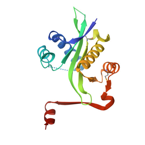
Crystal structure of Homo sapiens thialysine Nepsilon-acetyltransferase (HsSSAT2) in complex with acetyl coenzyme A.

Han, B.W.Bingman, C.A.Wesenberg, G.E.Phillips, G.N.

(2006) Proteins 64: 288-293


Macromolecules
Find similar proteins by:  (by identity cutoff)  |  3D Structure
Entity ID: 1
MoleculeChains Sequence LengthOrganismDetailsImage
Diamine acetyltransferase 2
A, B
170Homo sapiensMutation(s): 2 
Gene Names: SAT2SSAT2
EC: 2.3.1.57 (PDB Primary Data), 2.3.1 (UniProt)
UniProt & NIH Common Fund Data Resources
Find proteins for Q96F10 (Homo sapiens)
Explore Q96F10 
Go to UniProtKB:  Q96F10
PHAROS:  Q96F10
GTEx:  ENSG00000141504 
Entity Groups  
Sequence Clusters30% Identity50% Identity70% Identity90% Identity95% Identity100% Identity
UniProt GroupQ96F10
Sequence Annotations
Expand
  • Reference Sequence
Small Molecules
Ligands 1 Unique
IDChains Name / Formula / InChI Key2D Diagram3D Interactions
ACO
Query on ACO

Download Ideal Coordinates CCD File 
C [auth A]ACETYL COENZYME *A
C23 H38 N7 O17 P3 S
ZSLZBFCDCINBPY-ZSJPKINUSA-N
Modified Residues  1 Unique
IDChains TypeFormula2D DiagramParent
MSE
Query on MSE
A, B
L-PEPTIDE LINKINGC5 H11 N O2 SeMET
Experimental Data & Validation

Experimental Data

  • Method: X-RAY DIFFRACTION
  • Resolution: 1.84 Å
  • R-Value Free:  0.249 (Depositor), 0.247 (DCC) 
  • R-Value Work:  0.204 (Depositor), 0.204 (DCC) 
  • R-Value Observed: 0.207 (Depositor) 
Space Group: P 21 21 21
Unit Cell:
Length ( Å )Angle ( ˚ )
a = 54.583α = 90
b = 83.621β = 90
c = 87.548γ = 90
Software Package:
Software NamePurpose
DENZOdata reduction
SCALEPACKdata scaling
SHARPphasing
DMphasing
REFMACrefinement
PDB_EXTRACTdata extraction
SOLOMONphasing
ARP/wARPmodel building

Structure Validation

View Full Validation Report


Ligand Structure Quality Assessment 

Created with Raphaël 2.3.0Worse 01 BetterLigand structure goodness of fit to experimental dataBest fitted ACOClick on this verticalbar to view details

Entry History 

Revision History  (Full details and data files)

  • Version 1.0: 2005-11-01
    Type: Initial release
  • Version 1.1: 2008-05-01
    Changes: Version format compliance
  • Version 1.2: 2011-07-13
    Changes: Version format compliance
  • Version 1.3: 2017-10-18
    Changes: Refinement description
  • Version 1.4: 2024-10-30
    Changes: Data collection, Database references, Derived calculations, Structure summary