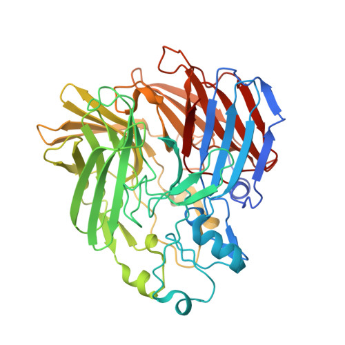
2BIW

Crystal structure of apocarotenoid cleavage oxygenase from Synechocystis, native enzyme


Experimental Data Snapshot

  • Method: X-RAY DIFFRACTION
  • Resolution: 2.39 Å
  • R-Value Free: 0.224 
  • R-Value Work: 0.180 
  • R-Value Observed: 0.182 

Starting Model: other
View more details

wwPDB Validation   3D Report Full Report


Ligand Structure Quality Assessment 


This is version 1.2 of the entry. See complete history


Literature

The Structure of a Retinal-Forming Carotenoid Oxygenase

Kloer, D.P.Ruch, S.Al-Babili, S.Beyer, P.Schulz, G.E.

(2005) Science 308: 267

  • DOI: https://doi.org/10.1126/science.1108965
  • Primary Citation of Related Structures:  
    2BIW, 2BIX

  • PubMed Abstract: 

    Enzymes that produce retinal and related apocarotenoids constitute a sequence- and thus structure-related family, a member of which was analyzed by x-ray diffraction. This member is an oxygenase and contains an Fe2+-4-His arrangement at the axis of a seven-bladed beta-propeller chain fold covered by a dome formed by six large loops. The Fe2+ is accessible through a long nonpolar tunnel that holds a carotenoid derivative in one of the crystals. On binding, three consecutive double bonds of this carotenoid changed from a straight all-trans to a cranked cis-trans-cis conformation. The remaining trans bond is located at the dioxygen-ligated Fe2+ and cleaved by oxygen.


  • Organizational Affiliation

    Institut für Organische Chemie und Biochemie, Albert-Ludwigs-Universität, Albertstrasse 21, 79104 Freiburg im Breisgau, Germany.


Macromolecules
Find similar proteins by:  (by identity cutoff)  |  3D Structure
Entity ID: 1
MoleculeChains Sequence LengthOrganismDetailsImage
APOCAROTENOID-CLEAVING OXYGENASE
A, B, C, D
490Synechocystis sp. PCC 6803Mutation(s): 0 
EC: 1.13.11.75
UniProt
Find proteins for P74334 (Synechocystis sp. (strain ATCC 27184 / PCC 6803 / Kazusa))
Explore P74334 
Go to UniProtKB:  P74334
Entity Groups  
Sequence Clusters30% Identity50% Identity70% Identity90% Identity95% Identity100% Identity
UniProt GroupP74334
Sequence Annotations
Expand
  • Reference Sequence
Small Molecules
Ligands 2 Unique
IDChains Name / Formula / InChI Key2D Diagram3D Interactions
3ON
Query on 3ON

Download Ideal Coordinates CCD File 
E [auth A],
G [auth B],
I [auth C],
K [auth D]
(3R)-3-HYDROXY-8'-APOCAROTENOL
C30 H42 O2
FNAJVVMDXCOSFY-VFGOXHQXSA-N
FE
Query on FE

Download Ideal Coordinates CCD File 
F [auth A],
H [auth B],
J [auth C],
L [auth D]
FE (III) ION
Fe
VTLYFUHAOXGGBS-UHFFFAOYSA-N
Experimental Data & Validation

Experimental Data

  • Method: X-RAY DIFFRACTION
  • Resolution: 2.39 Å
  • R-Value Free: 0.224 
  • R-Value Work: 0.180 
  • R-Value Observed: 0.182 
  • Space Group: P 21 21 21
Unit Cell:
Length ( Å )Angle ( ˚ )
a = 119.046α = 90
b = 125.278β = 90
c = 203.086γ = 90
Software Package:
Software NamePurpose
REFMACrefinement
XDSdata reduction
XSCALEdata scaling
MOLREPphasing

Structure Validation

View Full Validation Report


Ligand Structure Quality Assessment 


Entry History 

Deposition Data

Revision History  (Full details and data files)

  • Version 1.0: 2005-04-14
    Type: Initial release
  • Version 1.1: 2011-07-13
    Changes: Advisory, Version format compliance
  • Version 1.2: 2024-05-01
    Changes: Data collection, Database references, Derived calculations, Other, Refinement description