Structures of dimeric nonstandard nucleotide triphosphate pyrophosphatase from Pyrococcus horikoshii OT3: functional significance of interprotomer conformational changes
Lokanath, N.K., Pampa, K.J., Takio, K., Kunishima, N.(2008) J Mol Biol 375: 1013-1025
- PubMed: 18062990
- DOI: https://doi.org/10.1016/j.jmb.2007.11.018
- Primary Citation of Related Structures:
1V7R, 2DVN, 2DVO, 2DVP, 2ZTI - PubMed Abstract:
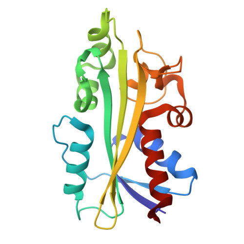
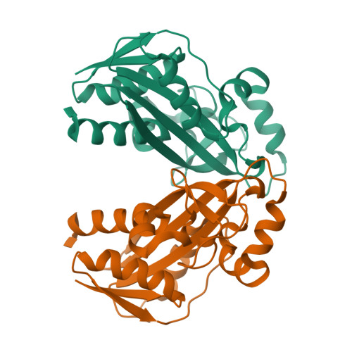
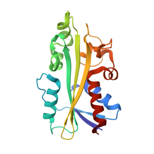
Nonstandard nucleotide triphosphate pyrophosphatase (NTPase) can efficiently hydrolyze nonstandard purine nucleotides in the presence of divalent cations. The crystal structures of the NTPase from Pyrococcus horikoshii OT3 (PhNTPase) have been determined in two unliganded forms and in three liganded forms with inosine 5'-monophosphate (IMP), ITP and Mn(2+), which visualize the recognition of these ligands unambiguously. The overall structure of PhNTPase is similar to that of previously reported crystal structures of the NTPase from Methanococcus jannaschii and the human ITPase. They share a similar protomer folding with two domains and a similar homodimeric quaternary structure. The dimeric interface of NTPase is well conserved, and the dimeric state might be important to constitute the active site of this enzyme. A conformational analysis of the five snapshots of PhNTPase structures using the multiple superposition method reveals that IMP, ITP and Mn(2+) bind to the active site without inducing large local conformational changes, indicating that a combination of interdomain and interprotomer rigid-body shifts mainly describes the conformational change of PhNTPase. The interdomain and interprotomer conformations of the ITP liganded form are essentially the same as those observed in the unliganded form 1, indicating that ITP binding to PhNTPase in solution may follow the selection mode in which ITP binds to the subunit that happens to be in the conformation observed in the unliganded form 1. In contrast to the human ITPase inducing a large domain closure upon ITP binding, the interdomain active site cleft is generally closed in PhNTPase and only the IMP binding form shows a remarkable domain opening by 14 degrees only in the B subunit. The interprotomer rigid-body rotation of PhNTPase has a tendency to keep the dimeric 2-fold symmetry, which is also true in human ITPase, thereby suggesting its relevance to the positive cooperativity of the dimeric NTPases. The exception of this rule is observed in the IMP liganded form in which the dimeric 2-fold symmetry is broken by a 3 degrees interprotomer rotation in an unusual direction. A combination of the exceptional interdomain and interprotomer relocations is most likely the reason for the observed asymmetric IMP binding that might be necessary for PhNTPase to release the reaction product IMP.
Organizational Affiliation:
Advanced Protein Crystallography Research Group, RIKEN SPring-8 Center, Harima Institute, 1-1-1 Kouto, Sayo-cho, Sayo-gun, Hyogo 679-5148, Japan.


