2ELB | pdb_00002elb

Crystal Structure of the BAR-PH domain of human APPL1


Experimental Data Snapshot

  • Method: X-RAY DIFFRACTION
  • Resolution: 2.60 Å
  • R-Value Free: 
    0.327 (Depositor), 0.335 (DCC) 
  • R-Value Work: 
    0.236 (Depositor), 0.247 (DCC) 
  • R-Value Observed: 
    0.236 (Depositor) 

wwPDB Validation 3D Report Full Report

Validation slider image for 2ELB

This is version 1.3 of the entry. See complete history

Literature

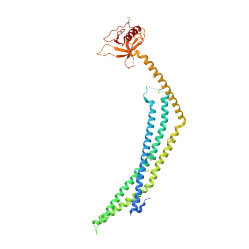
Crystal Structures of the BAR-PH and PTB Domains of Human APPL1

Li, J.Mao, X.Dong, L.Q.Liu, F.Tong, L.

(2007) Structure 15: 525-533

  • DOI: https://doi.org/10.1016/j.str.2007.03.011
  • Primary Citation Related Structures: 
    2ELA, 2ELB

  • PubMed Abstract: 

    APPL1 interacts with adiponectin receptors and other important signaling molecules. It contains a BAR and a PH domain near its N terminus, and the two domains may function as a unit (BAR-PH domain). We report here the crystal structures of the BAR-PH and PTB domains of human APPL1. The structures reveal novel features for BAR domain dimerization and for the interactions between the BAR and PH domains. The BAR domain dimer of APPL1 contains two four-helical bundles, whereas other BAR domain dimers have only three helices in each bundle. The PH domain is located at the opposite ends of the BAR domain dimer. Yeast two-hybrid assays confirm the interactions between the BAR and PH domains. Lipid binding assays show that the BAR, PH, and PTB domains can bind phospholipids. The ability of APPL1 to interact with multiple signaling molecules and phospholipids supports an important role for this adaptor in cell signaling.


  • Organizational Affiliation
    • Department of Biological Sciences, Columbia University, New York, NY 10027, USA.

Macromolecule Content 

  • Total Structure Weight: 45.99 kDa 
  • Atom Count: 2,834 
  • Modeled Residue Count: 351 
  • Deposited Residue Count: 396 
  • Unique protein chains: 1

Macromolecules

Find similar proteins by:|  3D Structure
Entity ID: 1
MoleculeChains  Sequence LengthOrganismDetailsImage
Adapter protein containing PH domain, PTB domain and leucine zipper motif 1396Homo sapiensMutation(s): 15 
UniProt & NIH Common Fund Data Resources
Find proteins for Q9UKG1 (Homo sapiens)
Explore Q9UKG1 
Go to UniProtKB:  Q9UKG1
PHAROS:  Q9UKG1
GTEx:  ENSG00000157500 
Entity Groups
Sequence Clusters30% Identity50% Identity70% Identity90% Identity95% Identity100% Identity
UniProt GroupQ9UKG1
Sequence Annotations
Expand
Reference Sequence

Small Molecules

Modified Residues  1 Unique
IDChains TypeFormula2D DiagramParent
MSE
Query on MSE
A
L-PEPTIDE LINKINGC5 H11 N O2 SeMET

Experimental Data & Validation

Experimental Data

  • Method: X-RAY DIFFRACTION
  • Resolution: 2.60 Å
  • R-Value Free:  0.327 (Depositor), 0.335 (DCC) 
  • R-Value Work:  0.236 (Depositor), 0.247 (DCC) 
  • R-Value Observed: 0.236 (Depositor) 
Space Group: P 21 21 2
Unit Cell:
Length ( Å )Angle ( ˚ )
a = 100.568α = 90
b = 104.126β = 90
c = 37.074γ = 90
Software Package:
Software NamePurpose
CNSrefinement
ADSCdata collection
DENZOdata reduction
SCALEPACKdata scaling
SnBphasing

Structure Validation

View Full Validation Report


Entry History 

Deposition Data

Revision History  (Full details and data files)

  • Version 1.0: 2007-05-29
    Type: Initial release
  • Version 1.1: 2008-04-30
    Changes: Version format compliance
  • Version 1.2: 2011-07-13
    Changes: Derived calculations, Version format compliance
  • Version 1.3: 2024-11-06
    Changes: Data collection, Database references, Derived calculations, Structure summary