


Crystal structure of human ribokinase
Rabeh, W.M., Tempel, W., Nedyalkova, L., Arrowsmith, C., Edwards, A., Sundstrom, M., Weigelt, J., Bochkarev, A., Park, H., Structural Genomics Consortium (SGC)To be published.
Experimental Data Snapshot
Starting Models: experimental
View more details
Entity ID: 1 | |||||
|---|---|---|---|---|---|
| Molecule | Chains | Sequence Length | Organism | Details | Image |
| Ribokinase | 331 | Homo sapiens | Mutation(s): 0 Gene Names: RBKS, RBSK EC: 2.7.1.15 | 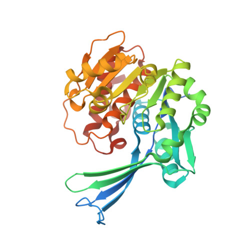 | |
UniProt & NIH Common Fund Data Resources | |||||
PHAROS: Q9H477 GTEx: ENSG00000171174 | |||||
Entity Groups | |||||
| Sequence Clusters | 30% Identity50% Identity70% Identity90% Identity95% Identity100% Identity | ||||
| UniProt Group | Q9H477 | ||||
Sequence AnnotationsExpand | |||||
Reference Sequence | |||||
| Ligands 4 Unique | |||||
|---|---|---|---|---|---|
| ID | Chains | Name / Formula / InChI Key | 2D Diagram | 3D Interactions | |
| ADP Download:Ideal Coordinates CCD File | E [auth A], N [auth B] | ADENOSINE-5'-DIPHOSPHATE C10 H15 N5 O10 P2 XTWYTFMLZFPYCI-KQYNXXCUSA-N |  | ||
| MG Download:Ideal Coordinates CCD File | D [auth A], M [auth B] | MAGNESIUM ION Mg JLVVSXFLKOJNIY-UHFFFAOYSA-N |  | ||
| NA Download:Ideal Coordinates CCD File | C [auth A], L [auth B] | SODIUM ION Na FKNQFGJONOIPTF-UHFFFAOYSA-N |  | ||
| UNX Download:Ideal Coordinates CCD File | F [auth A] G [auth A] H [auth A] I [auth A] J [auth A] | UNKNOWN ATOM OR ION X |  | ||
| Length ( Å ) | Angle ( ˚ ) |
|---|---|
| a = 45.53 | α = 90 |
| b = 72.978 | β = 91.08 |
| c = 90.958 | γ = 90 |
| Software Name | Purpose |
|---|---|
| DENZO | data reduction |
| SCALEPACK | data scaling |
| PHASER | phasing |
| DM | phasing |
| REFMAC | refinement |
| PDB_EXTRACT | data extraction |


