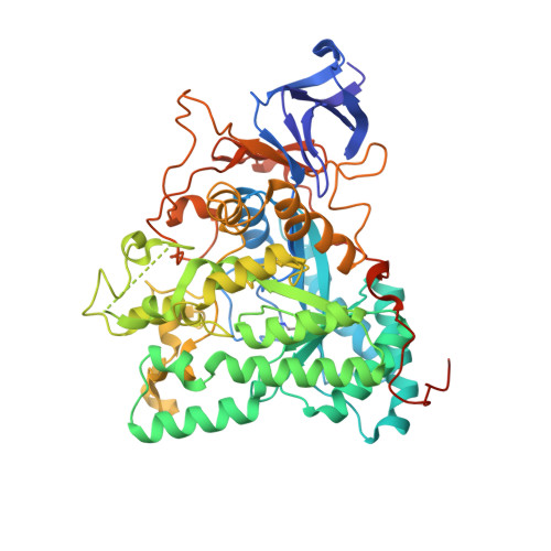
2FVK

Crystal structure of dihydropyrimidinase from Saccharomyces kluyveri in complex with the substrate dihydrouracil


Experimental Data Snapshot

  • Method: X-RAY DIFFRACTION
  • Resolution: 2.40 Å
  • R-Value Free: 0.234 
  • R-Value Work: 0.178 
  • R-Value Observed: 0.181 

Starting Model: experimental
View more details

wwPDB Validation   3D Report Full Report


This is version 1.4 of the entry. See complete history


Literature

The Crystal Structures of Dihydropyrimidinases Reaffirm the Close Relationship between Cyclic Amidohydrolases and Explain Their Substrate Specificity.

Lohkamp, B.Andersen, B.Piskur, J.Dobritzsch, D.

(2006) J Biol Chem 281: 13762-13776

  • DOI: https://doi.org/10.1074/jbc.M513266200
  • Primary Citation of Related Structures:  
    2FTW, 2FTY, 2FVK, 2FVM

  • PubMed Abstract: 

    In eukaryotes, dihydropyrimidinase catalyzes the second step of the reductive pyrimidine degradation, the reversible hydrolytic ring opening of dihydropyrimidines. Here we describe the three-dimensional structures of dihydropyrimidinase from two eukaryotes, the yeast Saccharomyces kluyveri and the slime mold Dictyostelium discoideum, determined and refined to 2.4 and 2.05 angstroms, respectively. Both enzymes have a (beta/alpha)8-barrel structural core embedding the catalytic di-zinc center, which is accompanied by a smaller beta-sandwich domain. Despite loop-forming insertions in the sequence of the yeast enzyme, the overall structures and architectures of the active sites of the dihydropyrimidinases are strikingly similar to each other, as well as to those of hydantoinases, dihydroorotases, and other members of the amidohydrolase superfamily of enzymes. However, formation of the physiologically relevant tetramer shows subtle but nonetheless significant differences. The extension of one of the sheets of the beta-sandwich domain across a subunit-subunit interface in yeast dihydropyrimidinase underlines its closer evolutionary relationship to hydantoinases, whereas the slime mold enzyme shows higher similarity to the noncatalytic collapsin-response mediator proteins involved in neuron development. Catalysis is expected to follow a dihydroorotase-like mechanism but in the opposite direction and with a different substrate. Complexes with dihydrouracil and N-carbamyl-beta-alanine obtained for the yeast dihydropyrimidinase reveal the mode of substrate and product binding and allow conclusions about what determines substrate specificity, stereoselectivity, and the reaction direction among cyclic amidohydrolases.


  • Organizational Affiliation

    Department of Medical Biochemistry and Biophysics, Karolinska Institutet, SE-17177 Stockholm, Sweden.


Macromolecules
Find similar proteins by:  (by identity cutoff)  |  3D Structure
Entity ID: 1
MoleculeChains Sequence LengthOrganismDetailsImage
dihydropyrimidinase
A, B, C, D
559Lachancea kluyveriMutation(s): 1 
Gene Names: Pyd2
EC: 3.5.2.2
UniProt
Find proteins for Q9P903 (Lachancea kluyveri (strain ATCC 58438 / CBS 3082 / BCRC 21498 / NBRC 1685 / JCM 7257 / NCYC 543 / NRRL Y-12651))
Explore Q9P903 
Go to UniProtKB:  Q9P903
Entity Groups  
Sequence Clusters30% Identity50% Identity70% Identity90% Identity95% Identity100% Identity
UniProt GroupQ9P903
Sequence Annotations
Expand
  • Reference Sequence
Small Molecules
Ligands 2 Unique
IDChains Name / Formula / InChI Key2D Diagram3D Interactions
DUC
Query on DUC

Download Ideal Coordinates CCD File 
G [auth A],
J [auth B],
M [auth C],
P [auth D]
DIHYDROPYRIMIDINE-2,4(1H,3H)-DIONE
C4 H6 N2 O2
OIVLITBTBDPEFK-UHFFFAOYSA-N
ZN
Query on ZN

Download Ideal Coordinates CCD File 
E [auth A]
F [auth A]
H [auth B]
I [auth B]
K [auth C]
E [auth A],
F [auth A],
H [auth B],
I [auth B],
K [auth C],
L [auth C],
N [auth D],
O [auth D]
ZINC ION
Zn
PTFCDOFLOPIGGS-UHFFFAOYSA-N
Modified Residues  1 Unique
IDChains TypeFormula2D DiagramParent
KCX
Query on KCX
A, B, C, D
L-PEPTIDE LINKINGC7 H14 N2 O4LYS
Experimental Data & Validation

Experimental Data

  • Method: X-RAY DIFFRACTION
  • Resolution: 2.40 Å
  • R-Value Free: 0.234 
  • R-Value Work: 0.178 
  • R-Value Observed: 0.181 
  • Space Group: P 1 21 1
Unit Cell:
Length ( Å )Angle ( ˚ )
a = 90.13α = 90
b = 71.6β = 91.4
c = 161.89γ = 90
Software Package:
Software NamePurpose
REFMACrefinement
MOSFLMdata reduction
CCP4data scaling

Structure Validation

View Full Validation Report


Entry History 

Deposition Data

Revision History  (Full details and data files)

  • Version 1.0: 2006-03-14
    Type: Initial release
  • Version 1.1: 2008-05-01
    Changes: Version format compliance
  • Version 1.2: 2011-07-13
    Changes: Advisory, Version format compliance
  • Version 1.3: 2023-10-25
    Changes: Data collection, Database references, Derived calculations, Refinement description
  • Version 1.4: 2023-11-15
    Changes: Data collection