2GSK | pdb_00002gsk

Structure of the BtuB:TonB Complex


Experimental Data Snapshot

  • Method: X-RAY DIFFRACTION
  • Resolution: 2.10 Å
  • R-Value Free: 
    0.251 (Depositor), 0.252 (DCC) 
  • R-Value Work: 
    0.196 (Depositor), 0.200 (DCC) 
  • R-Value Observed: 
    0.197 (Depositor) 

Starting Model: experimental
View more details

wwPDB Validation 3D Report Full Report

Validation slider image for 2GSK

Ligand Structure Quality Assessment 


This is version 2.1 of the entry. See complete history

Literature

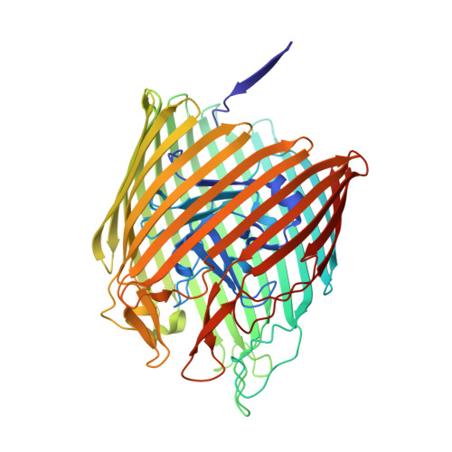
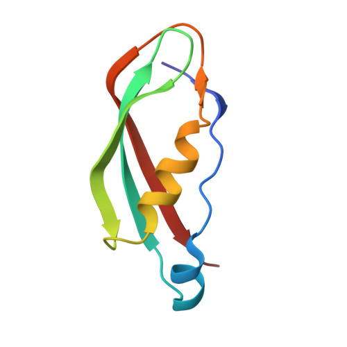
Outer membrane active transport: structure of the BtuB:TonB complex

Shultis, D.D.Purdy, M.D.Banchs, C.N.Wiener, M.C.

(2006) Science 312: 1396-1399

  • DOI: https://doi.org/10.1126/science.1127694
  • Primary Citation Related Structures: 
    2GSK

  • PubMed Abstract: 

    In Gram-negative bacteria, the import of essential micronutrients across the outer membrane requires a transporter, an electrochemical gradient of protons across the inner membrane, and an inner membrane protein complex (ExbB, ExbD, TonB) that couples the proton-motive force to the outer membrane transporter. The inner membrane protein TonB binds directly to a conserved region, called the Ton-box, of the transporter. We solved the structure of the cobalamin transporter BtuB in complex with the C-terminal domain of TonB. In contrast to its conformations in the absence of TonB, the Ton-box forms a beta strand that is recruited to the existing beta sheet of TonB, which is consistent with a mechanical pulling model of transport.


  • Organizational Affiliation
    • Department of Molecular Physiology and Biological Physics, University of Virginia, Charlottesville, VA 22908, USA.

Macromolecule Content 

  • Total Structure Weight: 78.07 kDa 
  • Atom Count: 5,658 
  • Modeled Residue Count: 671 
  • Deposited Residue Count: 671 
  • Unique protein chains: 2

Macromolecules

Find similar proteins by:|  3D Structure
Entity ID: 1
MoleculeChains  Sequence LengthOrganismDetailsImage
Vitamin B12 transporter btuB590Escherichia coliMutation(s): 0 
Gene Names: btuBbfecerdcrC
Membrane Entity: Yes 
UniProt
Find proteins for P06129 (Escherichia coli (strain K12))
Explore P06129 
Go to UniProtKB:  P06129
Entity Groups
Sequence Clusters30% Identity50% Identity70% Identity90% Identity95% Identity100% Identity
UniProt GroupP06129
Sequence Annotations
Expand
Reference Sequence
Find similar proteins by:|  3D Structure
Entity ID: 2
MoleculeChains  Sequence LengthOrganismDetailsImage
protein TONB81Escherichia coliMutation(s): 0 
Gene Names: tonBexbA
Membrane Entity: Yes 
UniProt
Find proteins for P02929 (Escherichia coli (strain K12))
Explore P02929 
Go to UniProtKB:  P02929
Entity Groups
Sequence Clusters30% Identity50% Identity70% Identity90% Identity95% Identity100% Identity
UniProt GroupP02929
Sequence Annotations
Expand
Reference Sequence

Small Molecules

Ligands 5 Unique
IDChains Name / Formula / InChI Key2D Diagram3D Interactions
CNC

Query on CNC


Download:Ideal Coordinates CCD File
E [auth A]CYANOCOBALAMIN
C63 H89 Co N14 O14 P
SYZBZQWSWIJYAR-UVKKECPRSA-M
LDA

Query on LDA


Download:Ideal Coordinates CCD File
F [auth A],
G [auth A],
H [auth A],
I [auth A]
LAURYL DIMETHYLAMINE-N-OXIDE
C14 H31 N O
SYELZBGXAIXKHU-UHFFFAOYSA-N
OCT

Query on OCT


Download:Ideal Coordinates CCD File
J [auth A],
K [auth A],
L [auth A]
N-OCTANE
C8 H18
TVMXDCGIABBOFY-UHFFFAOYSA-N
HEX

Query on HEX


Download:Ideal Coordinates CCD File
M [auth A],
N [auth A]
HEXANE
C6 H14
VLKZOEOYAKHREP-UHFFFAOYSA-N
CA

Query on CA


Download:Ideal Coordinates CCD File
C [auth A],
D [auth A]
CALCIUM ION
Ca
BHPQYMZQTOCNFJ-UHFFFAOYSA-N

Experimental Data & Validation

Experimental Data

  • Method: X-RAY DIFFRACTION
  • Resolution: 2.10 Å
  • R-Value Free:  0.251 (Depositor), 0.252 (DCC) 
  • R-Value Work:  0.196 (Depositor), 0.200 (DCC) 
  • R-Value Observed: 0.197 (Depositor) 
Space Group: P 21 21 21
Unit Cell:
Length ( Å )Angle ( ˚ )
a = 74.34α = 90
b = 82.439β = 90
c = 122.632γ = 90
Software Package:
Software NamePurpose
MAR345data collection
SERGUIdata collection
DENZOdata reduction
SCALEPACKdata scaling
MOLREPphasing
REFMACrefinement

Structure Validation

View Full Validation Report


Ligand Structure Quality Assessment 


Entry History 

Deposition Data

Revision History  (Full details and data files)

  • Version 1.0: 2006-06-13
    Type: Initial release
  • Version 1.1: 2008-05-01
    Changes: Version format compliance
  • Version 1.2: 2011-07-13
    Changes: Advisory, Version format compliance
  • Version 1.3: 2017-10-18
    Changes: Refinement description
  • Version 1.4: 2017-11-01
    Changes: Refinement description
  • Version 2.0: 2021-08-18
    Changes: Data collection, Database references, Derived calculations, Non-polymer description, Structure summary
  • Version 2.1: 2023-08-30
    Changes: Data collection, Refinement description