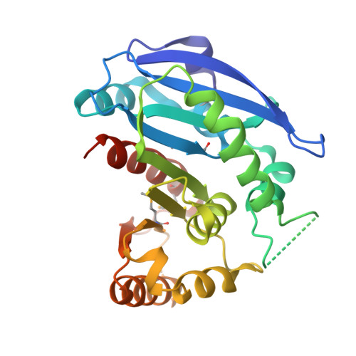
2GZR

Enterobactin and Salmochelin Hydrolase IroE


Experimental Data Snapshot

  • Method: X-RAY DIFFRACTION
  • Resolution: 2.30 Å
  • R-Value Free: 0.268 
  • R-Value Work: 0.203 

Starting Model: experimental
View more details

wwPDB Validation   3D Report Full Report


This is version 1.4 of the entry. See complete history


Literature

Structural Characterization of Enterobactin Hydrolase IroE.

Larsen, N.A.Lin, H.Wei, R.Fischbach, M.A.Walsh, C.T.

(2006) Biochemistry 45: 10184-10190

  • DOI: https://doi.org/10.1021/bi060950i
  • Primary Citation of Related Structures:  
    2GZR, 2GZS

  • PubMed Abstract: 

    The proliferation of many pathogenic bacteria is limited by the scarcity of soluble iron in their environment. Many of these bacteria scavenge iron by synthesizing and exporting small molecule siderophores that chelate iron. Iron-bound siderophores are subsequently imported for metabolic processing. Three related serine hydrolases have been characterized biochemically in this pathway: Fes, IroD, and IroE. Here, we report the crystal structure of IroE from uropathogenic Escherichia coli CFT073. The native structure and a complex with diisopropyl fluorophosphonate (DFP, a potent serine hydrolase inhibitor) were determined at 2.3 and 1.4 A resolution, respectively. IroE has the typical alpha/beta-hydrolase fold with an atypical catalytic dyad composed of Ser 189 and His 287. Mutation of either residue was detrimental to catalysis. In addition, rather than the typical oxyanion hole composed of backbone amides, IroE employs the atypical guanidinium moiety of Arg 130. Asp 90 anchors Arg 130 in the active site, and mutation of either residue was likewise detrimental to catalysis. We also compare the structure of IroE to the structure of Fes from Shigella flexneri (PDB entry 2B20). Both enzymes have similar active sites, but Fes has an additional amino-terminal lid domain. These lid domains are proposed to confer specificity to these related hydrolases.


  • Organizational Affiliation

    Department of Biological Chemistry and Molecular Pharmacology, Harvard Medical School, 240 Longwood Avenue, Boston, Massachusetts 02115-5702, USA.


Macromolecules
Find similar proteins by:  (by identity cutoff)  |  3D Structure
Entity ID: 1
MoleculeChains Sequence LengthOrganismDetailsImage
IroE protein278Escherichia coliMutation(s): 4 
Gene Names: iroE
UniProt
Find proteins for Q6KD95 (Escherichia coli)
Explore Q6KD95 
Go to UniProtKB:  Q6KD95
Entity Groups  
Sequence Clusters30% Identity50% Identity70% Identity90% Identity95% Identity100% Identity
UniProt GroupQ6KD95
Sequence Annotations
Expand
  • Reference Sequence
Small Molecules
Modified Residues  1 Unique
IDChains TypeFormula2D DiagramParent
MSE
Query on MSE
A
L-PEPTIDE LINKINGC5 H11 N O2 SeMET
Experimental Data & Validation

Experimental Data

  • Method: X-RAY DIFFRACTION
  • Resolution: 2.30 Å
  • R-Value Free: 0.268 
  • R-Value Work: 0.203 
  • Space Group: P 1
Unit Cell:
Length ( Å )Angle ( ˚ )
a = 36.22α = 111.469
b = 36.98β = 89.691
c = 44.208γ = 102.991
Software Package:
Software NamePurpose
CrystalCleardata collection
HKL-2000data reduction
MOLREPphasing
CNSrefinement
HKL-2000data scaling

Structure Validation

View Full Validation Report


Entry History 

Deposition Data

Revision History  (Full details and data files)

  • Version 1.0: 2006-09-05
    Type: Initial release
  • Version 1.1: 2008-05-01
    Changes: Version format compliance
  • Version 1.2: 2011-07-13
    Changes: Version format compliance
  • Version 1.3: 2023-08-30
    Changes: Data collection, Database references, Derived calculations, Refinement description
  • Version 1.4: 2023-11-15
    Changes: Data collection