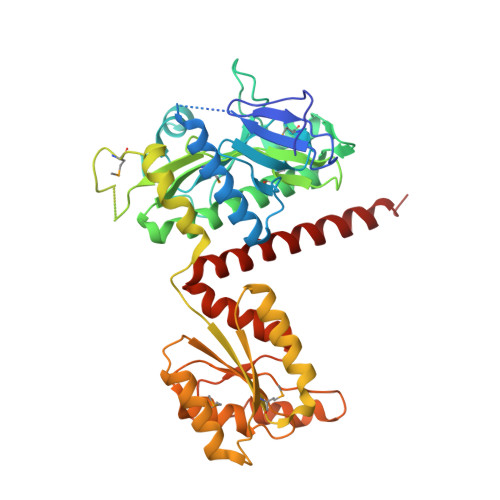
Octameric structure of the human bifunctional enzyme PAICS in purine biosynthesis.
Li, S.X., Tong, Y.P., Xie, X.C., Wang, Q.H., Zhou, H.N., Han, Y., Zhang, Z.Y., Gao, W., Li, S.G., Zhang, X.C., Bi, R.C.(2007) J Mol Biology 366: 1603-1614
- PubMed: 17224163
- DOI: https://doi.org/10.1016/j.jmb.2006.12.027
- Primary Citation of Related Structures:
2H31 - PubMed Abstract:
Phosphoribosylaminoimidazole carboxylase/phosphoribosylaminoimidazole succinocarboxamide synthetase (PAICS) is an important bifunctional enzyme in de novo purine biosynthesis in vertebrate with both 5-aminoimidazole ribonucleotide carboxylase (AIRc) and 4-(N-succinylcarboxamide)-5-aminoimidazole ribonucleotide synthetase (SAICARs) activities. It becomes an attractive target for rational anticancer drug design, since rapidly dividing cancer cells rely heavily on the purine de novo pathway for synthesis of adenine and guanine, whereas normal cells favor the salvage pathway. Here, we report the crystal structure of human PAICS, the first in the entire PAICS family, at 2.8 A resolution. It revealed that eight PAICS subunits, each composed of distinct AIRc and SAICARs domains, assemble a compact homo-octamer with an octameric-carboxylase core and four symmetric periphery dimers formed by synthetase domains. Based on structural comparison and functional complementation analyses, the active sites of SAICARs and AIRc were identified, including a putative substrate CO(2)-binding site. Furthermore, four symmetry-related, separate tunnel systems in the PAICS octamer were found that connect the active sites of AIRc and SAICARs. This study illustrated the octameric nature of the bifunctional enzyme. Each carboxylase active site is formed by structural elements from three AIRc domains, demonstrating that the octamer structure is essential for the carboxylation activity. Furthermore, the existence of the tunnel system implies a mechanism of intermediate channeling and suggests that the quaternary structure arrangement is crucial for effectively executing the sequential reactions. In addition, this study provides essential structural information for designing PAICS-specific inhibitors for use in cancer chemotherapy.
- Institute of Biophysics, Chinese Academy of Sciences, 15 Datun Road, Beijing 100101, China.
Organizational Affiliation:


