2H5A

Complex of the enzyme PMM/PGM with xylose 1-phosphate


Experimental Data Snapshot

  • Method: X-RAY DIFFRACTION
  • Resolution: 1.72 Å
  • R-Value Free: 
    0.213 (Depositor), 0.212 (DCC) 
  • R-Value Work: 
    0.176 (Depositor), 0.175 (DCC) 
  • R-Value Observed: 
    0.177 (Depositor) 

wwPDB Validation   3D Report Full Report


Ligand Structure Quality Assessment 

Created with Raphaël 2.3.0Worse 01 BetterLigand structure goodness of fit to experimental dataBest fitted X1PClick on this verticalbar to view details

This is version 1.5 of the entry. See complete history


Literature

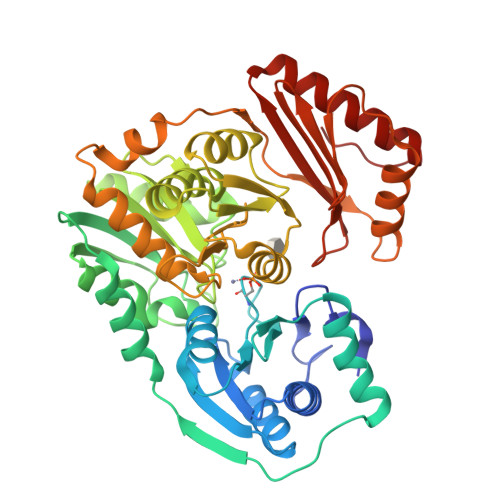
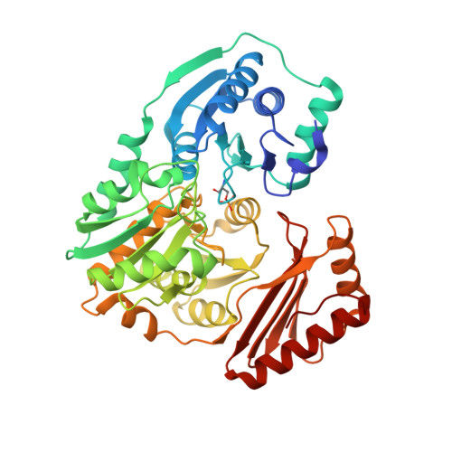
Complexes of the enzyme phosphomannomutase/phosphoglucomutase with a slow substrate and an inhibitor.

Regni, C.Shackelford, G.S.Beamer, L.J.

(2006) Acta Crystallogr Sect F Struct Biol Cryst Commun 62: 722-726

  • DOI: https://doi.org/10.1107/S1744309106025887
  • Primary Citation of Related Structures:  
    2H4L, 2H5A

  • PubMed Abstract: 

    Two complexes of the enzyme phosphomannomutase/phosphoglucomutase (PMM/PGM) from Pseudomonas aeruginosa with a slow substrate and with an inhibitor have been characterized by X-ray crystallography. Both ligands induce an interdomain rearrangement in the enzyme that creates a highly buried active site. Comparisons with enzyme-substrate complexes show that the inhibitor xylose 1-phosphate utilizes many of the previously observed enzyme-ligand interactions. In contrast, analysis of the ribose 1-phosphate complex reveals a combination of new and conserved enzyme-ligand interactions for binding. The ability of PMM/PGM to accommodate these two pentose phosphosugars in its active site may be relevant for future efforts towards inhibitor design.


  • Organizational Affiliation

    Department of Structural Biology, St Jude Children's Research Hospital, 332 North Lauderdale M/S 311, Memphis, TN 38105, USA.


Macromolecules
Find similar proteins by:  (by identity cutoff)  |  3D Structure
Entity ID: 1
MoleculeChains Sequence LengthOrganismDetailsImage
Phosphomannomutase/phosphoglucomutaseA [auth X]463Pseudomonas aeruginosaMutation(s): 1 
Gene Names: algC
EC: 5.4.2.8 (PDB Primary Data), 5.4.2.2 (PDB Primary Data)
UniProt
Find proteins for P26276 (Pseudomonas aeruginosa (strain ATCC 15692 / DSM 22644 / CIP 104116 / JCM 14847 / LMG 12228 / 1C / PRS 101 / PAO1))
Explore P26276 
Go to UniProtKB:  P26276
Entity Groups  
Sequence Clusters30% Identity50% Identity70% Identity90% Identity95% Identity100% Identity
UniProt GroupP26276
Sequence Annotations
Expand
  • Reference Sequence
Small Molecules
Modified Residues  1 Unique
IDChains TypeFormula2D DiagramParent
SEP
Query on SEP
A [auth X]L-PEPTIDE LINKINGC3 H8 N O6 PSER
Experimental Data & Validation

Experimental Data

  • Method: X-RAY DIFFRACTION
  • Resolution: 1.72 Å
  • R-Value Free:  0.213 (Depositor), 0.212 (DCC) 
  • R-Value Work:  0.176 (Depositor), 0.175 (DCC) 
  • R-Value Observed: 0.177 (Depositor) 
Space Group: P 21 21 21
Unit Cell:
Length ( Å )Angle ( ˚ )
a = 71.008α = 90
b = 74.551β = 90
c = 86.091γ = 90
Software Package:
Software NamePurpose
REFMACrefinement
HKL-2000data scaling

Structure Validation

View Full Validation Report


Ligand Structure Quality Assessment 

Created with Raphaël 2.3.0Worse 01 BetterLigand structure goodness of fit to experimental dataBest fitted X1PClick on this verticalbar to view details

Entry History 

Deposition Data

Revision History  (Full details and data files)

  • Version 1.0: 2006-08-08
    Type: Initial release
  • Version 1.1: 2008-05-01
    Changes: Version format compliance
  • Version 1.2: 2011-07-13
    Changes: Advisory, Version format compliance
  • Version 1.3: 2017-10-18
    Changes: Refinement description
  • Version 1.4: 2020-07-29
    Type: Remediation
    Reason: Carbohydrate remediation
    Changes: Database references, Derived calculations, Structure summary
  • Version 1.5: 2024-11-06
    Changes: Data collection, Database references, Structure summary