2HDF | pdb_00002hdf

Crystal structure of the Colicin I receptor Cir from E.coli


Experimental Data Snapshot

  • Method: X-RAY DIFFRACTION
  • Resolution: 2.65 Å
  • R-Value Free: 
    0.291 (Depositor), 0.288 (DCC) 
  • R-Value Work: 
    0.239 (Depositor) 
  • R-Value Observed: 
    0.241 (Depositor) 

wwPDB Validation 3D Report Full Report

Validation slider image for 2HDF

Ligand Structure Quality Assessment 


This is version 1.4 of the entry. See complete history

Literature

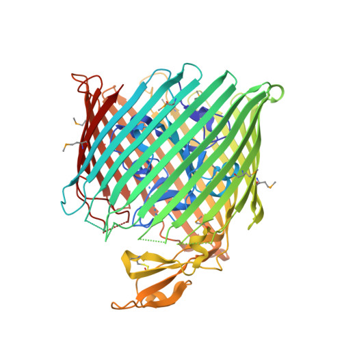
Structure of colicin I receptor bound to the R-domain of colicin Ia: implications for protein import.

Buchanan, S.K.Lukacik, P.Grizot, S.Ghirlando, R.Ali, M.M.Barnard, T.J.Jakes, K.S.Kienker, P.K.Esser, L.

(2007) EMBO J 26: 2594-2604

  • DOI: https://doi.org/10.1038/sj.emboj.7601693
  • Primary Citation Related Structures: 
    2HDF, 2HDI

  • PubMed Abstract: 

    Colicin Ia is a 69 kDa protein that kills susceptible Escherichia coli cells by binding to a specific receptor in the outer membrane, colicin I receptor (70 kDa), and subsequently translocating its channel forming domain across the periplasmic space, where it inserts into the inner membrane and forms a voltage-dependent ion channel. We determined crystal structures of colicin I receptor alone and in complex with the receptor binding domain of colicin Ia. The receptor undergoes large and unusual conformational changes upon colicin binding, opening at the cell surface and positioning the receptor binding domain of colicin Ia directly above it. We modelled the interaction with full-length colicin Ia to show that the channel forming domain is initially positioned 150 A above the cell surface. Functional data using full-length colicin Ia show that colicin I receptor is necessary for cell surface binding, and suggest that the receptor participates in translocation of colicin Ia across the outer membrane.


  • Organizational Affiliation
    • Laboratory of Molecular Biology, National Institute of Diabetes & Digestive & Kidney Diseases, National Institutes of Health, Bethesda, MD 20892, USA. skbuchan@helix.nih.gov

Macromolecule Content 

  • Total Structure Weight: 72.26 kDa 
  • Atom Count: 4,558 
  • Modeled Residue Count: 584 
  • Deposited Residue Count: 639 
  • Unique protein chains: 1

Macromolecules

Find similar proteins by:|  3D Structure
Entity ID: 1
MoleculeChains  Sequence LengthOrganismDetailsImage
Colicin I receptor639Escherichia coliMutation(s): 10 
Gene Names: cirA
Membrane Entity: Yes 
UniProt
Find proteins for P17315 (Escherichia coli (strain K12))
Explore P17315 
Go to UniProtKB:  P17315
Entity Groups
Sequence Clusters30% Identity50% Identity70% Identity90% Identity95% Identity100% Identity
UniProt GroupP17315
Sequence Annotations
Expand
Reference Sequence

Small Molecules

Experimental Data & Validation

Experimental Data

  • Method: X-RAY DIFFRACTION
  • Resolution: 2.65 Å
  • R-Value Free:  0.291 (Depositor), 0.288 (DCC) 
  • R-Value Work:  0.239 (Depositor) 
  • R-Value Observed: 0.241 (Depositor) 
Space Group: C 1 2 1
Unit Cell:
Length ( Å )Angle ( ˚ )
a = 90.593α = 90
b = 84.336β = 109.16
c = 99.458γ = 90
Software Package:
Software NamePurpose
REFMACrefinement
SHARPphasing

Structure Validation

View Full Validation Report


Ligand Structure Quality Assessment 


Entry History 

Deposition Data

Revision History  (Full details and data files)

  • Version 1.0: 2007-05-08
    Type: Initial release
  • Version 1.1: 2008-05-01
    Changes: Version format compliance
  • Version 1.2: 2011-07-13
    Changes: Advisory, Version format compliance
  • Version 1.3: 2021-10-20
    Changes: Database references, Derived calculations
  • Version 1.4: 2024-11-06
    Changes: Data collection, Structure summary