2IC8

Crystal structure of GlpG


Experimental Data Snapshot

  • Method: X-RAY DIFFRACTION
  • Resolution: 2.10 Å
  • R-Value Free: 
    0.252 (Depositor), 0.240 (DCC) 
  • R-Value Work: 
    0.236 (Depositor), 0.220 (DCC) 

wwPDB Validation   3D Report Full Report


Ligand Structure Quality Assessment 

Created with Raphaël 2.3.0Worse 01 BetterLigand structure goodness of fit to experimental dataBest fitted BNGClick on this verticalbar to view details

This is version 1.4 of the entry. See complete history


Literature

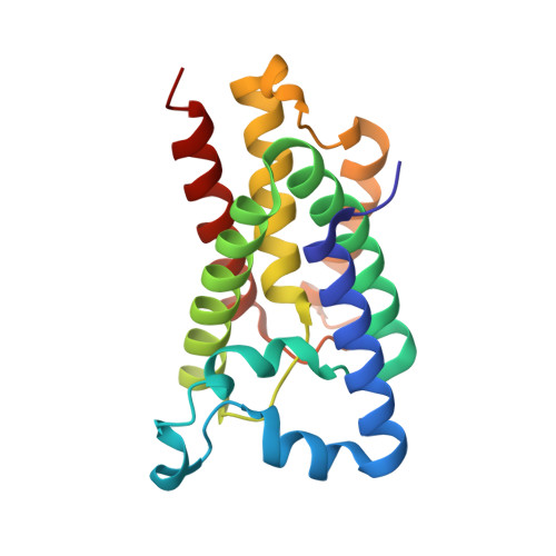
Crystal structure of a rhomboid family intramembrane protease.

Wang, Y.Zhang, Y.Ha, Y.

(2006) Nature 444: 179-180

  • DOI: https://doi.org/10.1038/nature05255
  • Primary Citation of Related Structures:  
    2IC8

  • PubMed Abstract: 

    Escherichia coli GlpG is an integral membrane protein that belongs to the widespread rhomboid protease family. Rhomboid proteases, like site-2 protease (S2P) and gamma-secretase, are unique in that they cleave the transmembrane domain of other membrane proteins. Here we describe the 2.1 A resolution crystal structure of the GlpG core domain. This structure contains six transmembrane segments. Residues previously shown to be involved in catalysis, including a Ser-His dyad, and several water molecules are found at the protein interior at a depth below the membrane surface. This putative active site is accessible by substrate through a large 'V-shaped' opening that faces laterally towards the lipid, but is blocked by a half-submerged loop structure. These observations indicate that, in intramembrane proteolysis, the scission of peptide bonds takes place within the hydrophobic environment of the membrane bilayer. The crystal structure also suggests a gating mechanism for GlpG that controls substrate access to its hydrophilic active site.


  • Organizational Affiliation

    Department of Pharmacology, Yale School of Medicine, 333 Cedar Street, New Haven, Connecticut 06520, USA.


Macromolecules
Find similar proteins by:  (by identity cutoff)  |  3D Structure
Entity ID: 1
MoleculeChains Sequence LengthOrganismDetailsImage
Protein glpG182Escherichia coliMutation(s): 0 
Gene Names: glpGb3424
EC: 3.4.21.105
Membrane Entity: Yes 
UniProt
Find proteins for P09391 (Escherichia coli (strain K12))
Explore P09391 
Go to UniProtKB:  P09391
Entity Groups  
Sequence Clusters30% Identity50% Identity70% Identity90% Identity95% Identity100% Identity
UniProt GroupP09391
Sequence Annotations
Expand
  • Reference Sequence
Small Molecules
Ligands 1 Unique
IDChains Name / Formula / InChI Key2D Diagram3D Interactions
BNG
Query on BNG

Download Ideal Coordinates CCD File 
B [auth A]
C [auth A]
D [auth A]
E [auth A]
F [auth A]
nonyl beta-D-glucopyranoside
C15 H30 O6
QFAPUKLCALRPLH-UXXRCYHCSA-N
Experimental Data & Validation

Experimental Data

  • Method: X-RAY DIFFRACTION
  • Resolution: 2.10 Å
  • R-Value Free:  0.252 (Depositor), 0.240 (DCC) 
  • R-Value Work:  0.236 (Depositor), 0.220 (DCC) 
Space Group: H 3 2
Unit Cell:
Length ( Å )Angle ( ˚ )
a = 110.822α = 90
b = 110.822β = 90
c = 127.611γ = 120
Software Package:
Software NamePurpose
CNSrefinement
PDB_EXTRACTdata extraction

Structure Validation

View Full Validation Report


Ligand Structure Quality Assessment 

Created with Raphaël 2.3.0Worse 01 BetterLigand structure goodness of fit to experimental dataBest fitted BNGClick on this verticalbar to view details

Entry History 

Deposition Data

  • Released Date: 2006-10-17 
  • Deposition Author(s): Ha, Y.

Revision History  (Full details and data files)

  • Version 1.0: 2006-10-17
    Type: Initial release
  • Version 1.1: 2008-05-01
    Changes: Version format compliance
  • Version 1.2: 2011-07-13
    Changes: Version format compliance
  • Version 1.3: 2020-07-29
    Type: Remediation
    Reason: Carbohydrate remediation
    Changes: Data collection, Derived calculations, Structure summary
  • Version 1.4: 2024-02-21
    Changes: Data collection, Database references, Structure summary