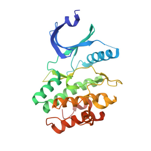
2IO6

Wee1 kinase complexed with inhibitor PD330961


Experimental Data Snapshot

  • Method: X-RAY DIFFRACTION
  • Resolution: 2.20 Å
  • R-Value Free: 0.273 
  • R-Value Work: 0.233 
  • R-Value Observed: 0.235 

Starting Model: experimental
View more details

wwPDB Validation   3D Report Full Report


Ligand Structure Quality Assessment 


This is version 1.2 of the entry. See complete history


Literature

Synthesis and structure-activity relationships of N-6 substituted analogues of 9-hydroxy-4-phenylpyrrolo[3,4-c]carbazole-1,3(2H,6H)-diones as inhibitors of Wee1 and Chk1 checkpoint kinases.

Smaill, J.B.Baker, E.N.Booth, R.J.Bridges, A.J.Dickson, J.M.Dobrusin, E.M.Ivanovic, I.Kraker, A.J.Lee, H.H.Lunney, E.A.Ortwine, D.F.Palmer, B.D.Quin, J.Squire, C.J.Thompson, A.M.Denny, W.A.

(2008) Eur J Med Chem 43: 1276-1296

  • DOI: https://doi.org/10.1016/j.ejmech.2007.07.016
  • Primary Citation of Related Structures:  
    2IN6, 2IO6

  • PubMed Abstract: 

    A series of N-6 substituted 9-hydroxy-4-phenylpyrrolo[3,4-c]carbazole-1,3(2H,6H)-diones were prepared from N-substituted (5-methoxyphenyl)ethenylindoles. The target compounds were tested for their ability to inhibit the G2/M cell cycle checkpoint kinases, Wee1 and Chk1. Analogues with neutral or cationic N-6 side chains were potent dual inhibitors. Acidic side chains provided potent (average IC(50) 0.057 microM) and selective (average ratio 223-fold) Wee1 inhibition. Co-crystal structures of inhibitors bound to Wee1 show that the pyrrolo[3,4-c]carbazole scaffold binds in the ATP-binding site, with N-6 substituents involved in H-bonding to conserved water molecules. HT-29 cells treated with doxorubicin and then target compounds demonstrate an active Cdc2/cyclin B complex, inhibition of the doxorubicin-induced phosphorylation of tyrosine 15 of Cdc2 and abrogation of the G2 checkpoint.


  • Organizational Affiliation

    Auckland Cancer Society Research Centre, School of Medical Sciences, The University of Auckland, Private Bag 92019, Auckland 1020, New Zealand. j.smaill@auckland.ac.nz


Macromolecules
Find similar proteins by:  (by identity cutoff)  |  3D Structure
Entity ID: 1
MoleculeChains Sequence LengthOrganismDetailsImage
Wee1-like protein kinase287Homo sapiensMutation(s): 0 
Gene Names: WEE1
EC: 2.7.10.2
UniProt & NIH Common Fund Data Resources
Find proteins for P30291 (Homo sapiens)
Explore P30291 
Go to UniProtKB:  P30291
PHAROS:  P30291
GTEx:  ENSG00000166483 
Entity Groups  
Sequence Clusters30% Identity50% Identity70% Identity90% Identity95% Identity100% Identity
UniProt GroupP30291
Sequence Annotations
Expand
  • Reference Sequence
Small Molecules
Ligands 1 Unique
IDChains Name / Formula / InChI Key2D Diagram3D Interactions
330
Query on 330

Download Ideal Coordinates CCD File 
B [auth A]9-HYDROXY-6-(3-HYDROXYPROPYL)-4-(2-METHOXYPHENYL)PYRROLO[3,4-C]CARBAZOLE-1,3(2H,6H)-DIONE
C24 H20 N2 O5
AOGOZJCRIFBTTN-UHFFFAOYSA-N
Binding Affinity Annotations 
IDSourceBinding Affinity
330 PDBBind:  2IO6 IC50: 30 (nM) from 1 assay(s)
BindingDB:  2IO6 IC50: 30 (nM) from 1 assay(s)
Experimental Data & Validation

Experimental Data

  • Method: X-RAY DIFFRACTION
  • Resolution: 2.20 Å
  • R-Value Free: 0.273 
  • R-Value Work: 0.233 
  • R-Value Observed: 0.235 
  • Space Group: P 41 21 2
Unit Cell:
Length ( Å )Angle ( ˚ )
a = 69.925α = 90
b = 69.925β = 90
c = 157.92γ = 90
Software Package:
Software NamePurpose
REFMACrefinement
HKL-2000data collection
HKL-2000data reduction
SCALEPACKdata scaling
AMoREphasing

Structure Validation

View Full Validation Report


Ligand Structure Quality Assessment 


Entry History 

Deposition Data

Revision History  (Full details and data files)

  • Version 1.0: 2007-09-18
    Type: Initial release
  • Version 1.1: 2011-07-13
    Changes: Version format compliance
  • Version 1.2: 2023-08-30
    Changes: Data collection, Database references, Derived calculations, Refinement description