2IYP | pdb_00002iyp

product rup


Experimental Data Snapshot

  • Method: X-RAY DIFFRACTION
  • Resolution: 2.79 Å
  • R-Value Free: 
    0.255 (Depositor), 0.254 (DCC) 
  • R-Value Work: 
    0.179 (Depositor), 0.179 (DCC) 
  • R-Value Observed: 
    0.182 (Depositor) 

wwPDB Validation 3D Report Full Report

Validation slider image for 2IYP

Ligand Structure Quality Assessment 


This is version 1.3 of the entry. See complete history

Literature

Crystal Structures of a Bacterial 6-Phosphogluconate Dehydrogenase Reveal Aspects of Specificity, Mechanism and Mode of Inhibition by Analogues of High-Energy Reaction Intermediates.

Sundaramoorthy, R.Iulek, J.Barrett, M.P.Bidet, O.Ruda, G.F.Gilbert, I.H.Hunter, W.N.

(2007) FEBS J 274: 275

  • DOI: https://doi.org/10.1111/j.1742-4658.2006.05585.x
  • Primary Citation Related Structures: 
    2IYO, 2IYP, 2IZ0, 2IZ1

  • PubMed Abstract: 

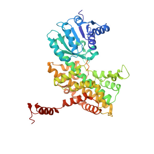
    Crystal structures of recombinant Lactococcus lactis 6-phosphogluconate dehydrogenase (LlPDH) in complex with substrate, cofactor, product and inhibitors have been determined. LlPDH shares significant sequence identity with the enzymes from sheep liver and the protozoan parasite Trypanosoma brucei for which structures have been reported. Comparisons indicate that the key residues in the active site are highly conserved, as are the interactions with the cofactor and the product ribulose 5-phosphate. However, there are differences in the conformation of the substrate 6-phosphogluconate which may reflect distinct states relevant to catalysis. Analysis of the complex formed with the potent inhibitor 4-phospho-d-erythronohydroxamic acid, suggests that this molecule does indeed mimic the high-energy intermediate state that it was designed to. The analysis also identified, as a contaminant by-product of the inhibitor synthesis, 4-phospho-d-erythronamide, which binds in similar fashion. LlPDH can now serve as a model system for structure-based inhibitor design targeting the enzyme from Trypanosoma species.


  • Organizational Affiliation
    • Division of Biological Chemistry and Molecular Microbiology, College of Life Sciences, University of Dundee, UK.

Macromolecule Content 

  • Total Structure Weight: 159.75 kDa 
  • Atom Count: 11,578 
  • Modeled Residue Count: 1,410 
  • Deposited Residue Count: 1,419 
  • Unique protein chains: 1

Macromolecules

Find similar proteins by:|  3D Structure
Entity ID: 1
MoleculeChains  Sequence LengthOrganismDetailsImage
6-PHOSPHOGLUCONATE DEHYDROGENASE, DECARBOXYLATING
A, B, C
473Lactococcus lactisMutation(s): 0 
EC: 1.1.1.44
UniProt
Find proteins for P96789 (Lactococcus lactis subsp. cremoris (strain MG1363))
Explore P96789 
Go to UniProtKB:  P96789
Entity Groups
Sequence Clusters30% Identity50% Identity70% Identity90% Identity95% Identity100% Identity
UniProt GroupP96789
Sequence Annotations
Expand
Reference Sequence

Experimental Data & Validation

Experimental Data

  • Method: X-RAY DIFFRACTION
  • Resolution: 2.79 Å
  • R-Value Free:  0.255 (Depositor), 0.254 (DCC) 
  • R-Value Work:  0.179 (Depositor), 0.179 (DCC) 
  • R-Value Observed: 0.182 (Depositor) 
Space Group: C 1 2 1
Unit Cell:
Length ( Å )Angle ( ˚ )
a = 71.062α = 90
b = 105.061β = 98.26
c = 240.484γ = 90
Software Package:
Software NamePurpose
REFMACrefinement
DENZOdata reduction
SCALEPACKdata scaling
PHASERphasing

Structure Validation

View Full Validation Report


Ligand Structure Quality Assessment 


Entry History 

Deposition Data

Revision History  (Full details and data files)

  • Version 1.0: 2007-01-23
    Type: Initial release
  • Version 1.1: 2011-05-08
    Changes: Version format compliance
  • Version 1.2: 2011-07-13
    Changes: Version format compliance
  • Version 1.3: 2024-05-08
    Changes: Data collection, Database references, Other