2J2Z | pdb_00002j2z

X-Ray Structure of the Chaperone PapD in complex with the Pilus terminator subunit PapH at 2.3 Angstrom resolution


Experimental Data Snapshot

  • Method: X-RAY DIFFRACTION
  • Resolution: 2.30 Å
  • R-Value Free: 
    0.240 (Depositor), 0.234 (DCC) 
  • R-Value Work: 
    0.209 (Depositor), 0.202 (DCC) 
  • R-Value Observed: 
    0.209 (Depositor) 

Starting Model: experimental
View more details

wwPDB Validation 3D Report Full Report

Validation slider image for 2J2Z

This is version 1.4 of the entry. See complete history

Literature

Molecular Mechanism of P Pilus Termination in Uropathogenic Escherichia Coli.

Verger, D.Miller, E.Remaut, H.Waksman, G.Hultgren, S.

(2006) EMBO Rep 7: 1228

  • DOI: https://doi.org/10.1038/sj.embor.7400833
  • Primary Citation Related Structures: 
    2J2Z

  • PubMed Abstract: 

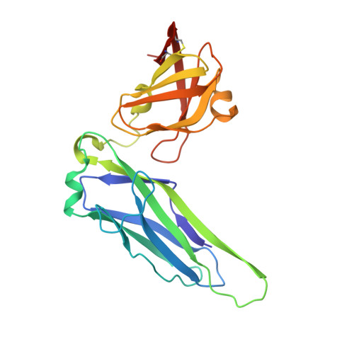
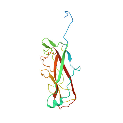
    P pili are important adhesive fibres that are assembled by the conserved chaperone-usher pathway. During pilus assembly, the subunits are incorporated into the growing fibre by the donor-strand exchange mechanism, whereby the beta-strand of the chaperone, which complements the incomplete immunoglobulin fold of each subunit, is displaced by the amino-terminal extension of an incoming subunit in a zip-in-zip-out exchange process that is initiated at the P5 pocket, an exposed hydrophobic pocket in the groove of the subunit. In vivo, termination of P pilus growth requires a specialized subunit, PapH. Here, we show that PapH is incorporated at the base of the growing pilus, where it is unable to undergo donor-strand exchange. This inability is not due to a stronger PapD-PapH interaction, but to a lack of a P5 initiator pocket in the PapH structure, suggesting that PapH terminates pilus growth because it is lacking the initiation point by which donor-strand exchange proceeds.


  • Organizational Affiliation
    • Institute of Structural Molecular Biology, University College London, Birkbeck College, Malet Street, London WC1E 7HX, UK.

Macromolecule Content 

  • Total Structure Weight: 45 kDa 
  • Atom Count: 3,134 
  • Modeled Residue Count: 367 
  • Deposited Residue Count: 391 
  • Unique protein chains: 2

Macromolecules

Find similar proteins by:|  3D Structure
Entity ID: 1
MoleculeChains  Sequence LengthOrganismDetailsImage
CHAPERONE PROTEIN PAPD218Escherichia coliMutation(s): 0 
UniProt
Find proteins for P15319 (Escherichia coli)
Explore P15319 
Go to UniProtKB:  P15319
Entity Groups
Sequence Clusters30% Identity50% Identity70% Identity90% Identity95% Identity100% Identity
UniProt GroupP15319
Sequence Annotations
Expand
Reference Sequence
Find similar proteins by:|  3D Structure
Entity ID: 2
MoleculeChains  Sequence LengthOrganismDetailsImage
PAP FIMBRIAL MINOR PILIN PROTEIN173Escherichia coliMutation(s): 0 
UniProt
Find proteins for P07111 (Escherichia coli)
Explore P07111 
Go to UniProtKB:  P07111
Entity Groups
Sequence Clusters30% Identity50% Identity70% Identity90% Identity95% Identity100% Identity
UniProt GroupP07111
Sequence Annotations
Expand
Reference Sequence

Small Molecules

Ligands 2 Unique
IDChains Name / Formula / InChI Key2D Diagram3D Interactions
SO4

Query on SO4


Download:Ideal Coordinates CCD File
C [auth A]
D [auth A]
E [auth A]
F [auth A]
G [auth A]
C [auth A],
D [auth A],
E [auth A],
F [auth A],
G [auth A],
H [auth A],
J [auth B],
K [auth B],
L [auth B],
M [auth B],
N [auth B]
SULFATE ION
O4 S
QAOWNCQODCNURD-UHFFFAOYSA-L
CO

Query on CO


Download:Ideal Coordinates CCD File
I [auth B]COBALT (II) ION
Co
XLJKHNWPARRRJB-UHFFFAOYSA-N

Experimental Data & Validation

Experimental Data

  • Method: X-RAY DIFFRACTION
  • Resolution: 2.30 Å
  • R-Value Free:  0.240 (Depositor), 0.234 (DCC) 
  • R-Value Work:  0.209 (Depositor), 0.202 (DCC) 
  • R-Value Observed: 0.209 (Depositor) 
Space Group: C 2 2 21
Unit Cell:
Length ( Å )Angle ( ˚ )
a = 103.46α = 90
b = 149.3β = 90
c = 82.84γ = 90
Software Package:
Software NamePurpose
CNSrefinement
MOSFLMdata reduction
SCALEPACKdata scaling
AMoREphasing

Structure Validation

View Full Validation Report


Entry History 

Deposition Data

Revision History  (Full details and data files)

  • Version 1.0: 2006-11-08
    Type: Initial release
  • Version 1.1: 2011-05-08
    Changes: Version format compliance
  • Version 1.2: 2011-07-13
    Changes: Version format compliance
  • Version 1.3: 2023-12-13
    Changes: Data collection, Database references, Derived calculations, Other, Refinement description
  • Version 1.4: 2024-11-06
    Changes: Structure summary