2JG3 | pdb_00002jg3

MtaqI with BAZ


Experimental Data Snapshot

  • Method: X-RAY DIFFRACTION
  • Resolution: 1.90 Å
  • R-Value Free: 
    0.226 (Depositor), 0.223 (DCC) 
  • R-Value Work: 
    0.185 (Depositor), 0.185 (DCC) 
  • R-Value Observed: 
    0.187 (Depositor) 

Starting Model: experimental
View more details

wwPDB Validation 3D Report Full Report

Validation slider image for 2JG3

Ligand Structure Quality Assessment 


This is version 1.4 of the entry. See complete history

Literature

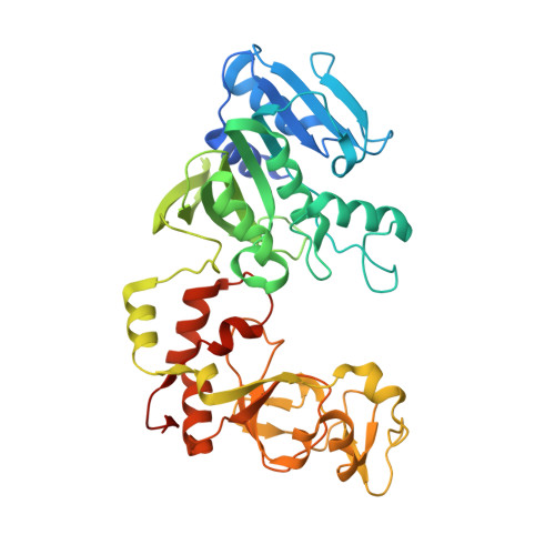
Quantitative Labeling of Long Plasmid DNA with Nanometer Precision.

Pljevaljcic, G.Schmidt, F.Scheidig, A.J.Lurz, R.Weinhold, E.

(2007) Chembiochem 8: 1516

Macromolecule Content 

  • Total Structure Weight: 109.54 kDa 
  • Atom Count: 7,895 
  • Modeled Residue Count: 824 
  • Deposited Residue Count: 882 
  • Unique protein chains: 1
  • Unique nucleic acid chains: 2

Macromolecules


Find similar proteins by:|  3D Structure
Entity ID: 1
MoleculeChains  Sequence LengthOrganismDetailsImage
MODIFICATION METHYLASE TAQI
A, D
421Thermus aquaticusMutation(s): 0 
EC: 2.1.1.72
UniProt
Find proteins for P14385 (Thermus aquaticus)
Explore P14385 
Go to UniProtKB:  P14385
Entity Groups
Sequence Clusters30% Identity50% Identity70% Identity90% Identity95% Identity100% Identity
UniProt GroupP14385
Sequence Annotations
Expand
Reference Sequence
Find similar nucleic acids by:  Sequence
Entity ID: 2
MoleculeChains LengthOrganismImage
5'-D(*GP*TP*TP*CP*GP*AP*TP*GP*TP*CP)-3'
B, E
10synthetic construct
Sequence Annotations
Expand
Reference Sequence
Find similar nucleic acids by:  Sequence
Entity ID: 3
MoleculeChains LengthOrganismImage
5'-D(*GP*AP*CP*AP*TP*CP*GP*6MAP*AP*CP)-3'
C, F
10synthetic construct
Sequence Annotations
Expand
Reference Sequence

Small Molecules

Ligands 3 Unique
IDChains Name / Formula / InChI Key2D Diagram3D Interactions
BA2

Query on BA2


Download:Ideal Coordinates CCD File
G [auth A],
L [auth D]
5'-DEOXY-5'-(ETHYLAMINO)-8-{[4-({5-[(3AS,4S,6AR)-2-OXOHEXAHYDRO-1H-THIENO[3,4-D]IMIDAZOL-4-YL]PENTANOYL}AMINO)BUTYL]AMINO}ADENOSINE
C26 H42 N10 O5 S
GZOCMSGHLSHCGU-DGWADGOTSA-N
GOL

Query on GOL


Download:Ideal Coordinates CCD File
H [auth A],
J [auth B]
GLYCEROL
C3 H8 O3
PEDCQBHIVMGVHV-UHFFFAOYSA-N
K

Query on K


Download:Ideal Coordinates CCD File
I [auth B],
K [auth D]
POTASSIUM ION
K
NPYPAHLBTDXSSS-UHFFFAOYSA-N

Experimental Data & Validation

Experimental Data

  • Method: X-RAY DIFFRACTION
  • Resolution: 1.90 Å
  • R-Value Free:  0.226 (Depositor), 0.223 (DCC) 
  • R-Value Work:  0.185 (Depositor), 0.185 (DCC) 
  • R-Value Observed: 0.187 (Depositor) 
Space Group: P 1 21 1
Unit Cell:
Length ( Å )Angle ( ˚ )
a = 59.404α = 90
b = 69.193β = 92.16
c = 114.39γ = 90
Software Package:
Software NamePurpose
REFMACrefinement
MARdata reduction
XDSdata scaling
MOLREPphasing

Structure Validation

View Full Validation Report


Ligand Structure Quality Assessment 


Entry History 

Deposition Data

Revision History  (Full details and data files)

  • Version 1.0: 2007-08-07
    Type: Initial release
  • Version 1.1: 2011-07-13
    Changes: Refinement description, Version format compliance
  • Version 1.2: 2019-01-30
    Changes: Data collection, Experimental preparation, Source and taxonomy
  • Version 1.3: 2019-02-06
    Changes: Data collection, Experimental preparation
  • Version 1.4: 2023-12-13
    Changes: Data collection, Database references, Derived calculations, Other, Refinement description