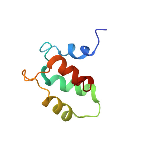
Solution structure of the Apo C-terminal domain of the Lethocerus F1 troponin C isoform.
De Nicola, G.F., Martin, S., Bullard, B., Pastore, A.(2010) Biochemistry 49: 1719-1726
- PubMed: 20104876 Search on PubMedSearch on PubMed Central
- DOI: https://doi.org/10.1021/bi902094w
- Primary Citation Related Structures:
2K2A - PubMed Abstract:
Muscle contraction is activated by two distinct mechanisms. One depends on the calcium influx, and the other is calcium-independent and activated by mechanical stress. A prototypical example of stretch activation is observed in insect muscles. In Lethocerus, a model system ideally suited for studying stretch activation, the two mechanisms seem to be under the control of different isoforms of troponin C (TnC), F1 and F2, which are responsible for stretch and calcium-dependent regulation, respectively. We have previously shown that F1 TnC is a typical collapsed dumbbell EF-hand protein that accommodates one calcium ion in its fourth EF-hand. When calcium loaded, the C-terminal domain of F1 TnC is in an open conformation which allows binding to troponin I. We have determined the solution structure of the isolated F1 TnC C-terminal domain in the absence of calcium and have compared it together with its dynamical properties with those of the calcium-loaded form. The domain is folded also in the absence of calcium and is in a closed conformation. Binding of a single calcium is sufficient to induce a modest but clear closed-to-open conformational transition and releases the conformational entropy observed in the calcium-free form. These results provide the first example of a TnC domain in which the presence of only one calcium ion is sufficient to induce a closed-to-open transition and clarify the role of calcium in stretch activation.
- Molecular Structure Division, National Institute for Medical Research, MRC, The Ridgeway, Mill Hill, London NW71AA, UK.
Organizational Affiliation: