2KC5 | pdb_00002kc5

Solution Structure of HybE from Escherichia coli


Experimental Data Snapshot

  • Method: SOLUTION NMR
  • Conformers Calculated: 100 
  • Conformers Submitted: 20 
  • Selection Criteria: structures with the lowest energy 

wwPDB Validation 3D Report Full Report

Validation slider image for 2KC5

This is version 1.2 of the entry. See complete history

Literature

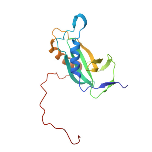
Solution structure of the Escherichia coli HybE reveals a novel fold

Shao, X.Lu, J.Hu, Y.Xia, B.Jin, C.

(2009) Proteins 75: 1051-1056

Macromolecule Content 

  • Total Structure Weight: 17.98 kDa 
  • Atom Count: 1,259 
  • Modeled Residue Count: 162 
  • Deposited Residue Count: 162 
  • Unique protein chains: 1

Macromolecules

Find similar proteins by:|  3D Structure
Entity ID: 1
MoleculeChains  Sequence LengthOrganismDetailsImage
Hydrogenase-2 operon protein hybE162Escherichia coli K-12Mutation(s): 0 
Gene Names: hybE
UniProt
Find proteins for P0AAN1 (Escherichia coli (strain K12))
Explore P0AAN1 
Go to UniProtKB:  P0AAN1
Entity Groups
Sequence Clusters30% Identity50% Identity70% Identity90% Identity95% Identity100% Identity
UniProt GroupP0AAN1
Sequence Annotations
Expand
Reference Sequence

Experimental Data & Validation

Experimental Data

  • Method: SOLUTION NMR
  • Conformers Calculated: 100 
  • Conformers Submitted: 20 
  • Selection Criteria: structures with the lowest energy 

Structure Validation

View Full Validation Report


Entry History 

Deposition Data

Revision History  (Full details and data files)

  • Version 1.0: 2009-10-20
    Type: Initial release
  • Version 1.1: 2011-07-13
    Changes: Source and taxonomy, Version format compliance
  • Version 1.2: 2024-05-01
    Changes: Data collection, Database references