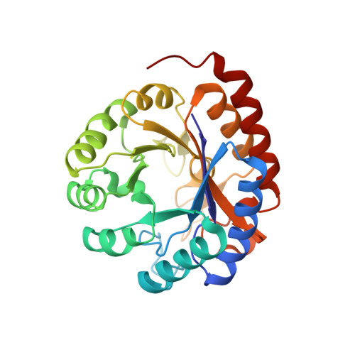
Structural and Mechanistic Changes along an Engineered Path from Metallo to Nonmetallo 3-Deoxy-d-manno-octulosonate 8-Phosphate Synthases.
Kona, F., Xu, X., Martin, P., Kuzmic, P., Gatti, D.L.(2007) Biochemistry 46: 4532-4544
- PubMed: 17381075
- DOI: https://doi.org/10.1021/bi6024879
- Primary Citation of Related Structures:
2EF9, 2NWR, 2NWS, 2NX1, 2NX3, 2NXG, 2NXH, 2NXI - PubMed Abstract:
There are two classes of KDO8P synthases characterized respectively by the presence or absence of a metal in the active site. The nonmetallo KDO8PS from Escherichia coli and the metallo KDO8PS from Aquifex aeolicus are the best characterized members of each class. All amino acid residues that make important contacts with the substrates are conserved in both enzymes with the exception of Pro-10, Cys-11, Ser-235, and Gln-237 of the A. aeolicus enzyme, which correspond respectively to Met-25, Asn-26, Pro-252, and Ala-254 in the E. coli enzyme. Interconversion between the two forms of KDO8P synthases can be achieved by substituting the metal-coordinating cysteine of metallo synthases with the corresponding asparagine of nonmetallo synthases, and vice versa. In this report we describe the structural changes elicited by the C11N mutation and by three combinations of mutations (P10M/C11N, C11N/S235P/Q237A, and P10M/C11N/S235P/Q237A) situated along possible evolutionary paths connecting the A. aeolicus and the E. coli enzyme. All four mutants are not capable of binding metal and lack the structural asymmetry among subunits with regard to substrate binding and conformation of the L7 loop, which is typical of A. aeolicus wild-type KDO8PS but is absent in the E. coli enzyme. Despite the lack of the active site metal, the mutant enzymes display levels of activity ranging from 46% to 24% of the wild type. With the sole exception of the quadruple mutant, metal loss does not affect the thermal stability of KDO8PS. The free energy of unfolding in water is also either unchanged or even increased in the mutant enzymes, suggesting that the primary role of the active site metal in A. aeolicus KDO8PS is not to increase the enzyme stability. In all four mutants A5P binding displaces a water molecule located on the si side of PEP. In particular, in the double and triple mutant, A5P binds with the aldehyde carbonyl in hydrogen bond distance of Asn-11, while in the wild type this functional group points away from Cys-11. This alternative conformation of A5P is likely to have functional significance as it resembles the conformation of the acyclic reaction intermediate, which is observed here for the first time in some of the active sites of the triple mutant. The direct visualization of this intermediate by X-ray crystallography confirms earlier mechanistic models of KDO8P synthesis. In particular, the configuration of the C2 chiral center of the intermediate supports a model of the reaction in nonmetallo KDO8PS, in which water attacks an oxocarbenium ion or PEP from the si side of C2. Several explanations are offered to reconcile this observation with the fact that no water molecule is observed at this position in the mutant enzymes in the presence of both PEP and A5P. Significant differences were observed between the wild-type and the mutant enzymes in the Km values for PEP and A5P and in the Kd values for inorganic phosphate and R5P. These differences may reflect an evolutionary adaptation of metallo and nonmetallo KDO8PS's to the cellular concentrations of these metabolites in their respective hosts.
- Department of Biochemistry and Molecular Biology, Wayne State University School of Medicine, Detroit, Michigan 48201, USA.
Organizational Affiliation:


