2PG7

Crystal Structure of Human Microsomal P450 2A6 N297Q/I300V


Experimental Data Snapshot

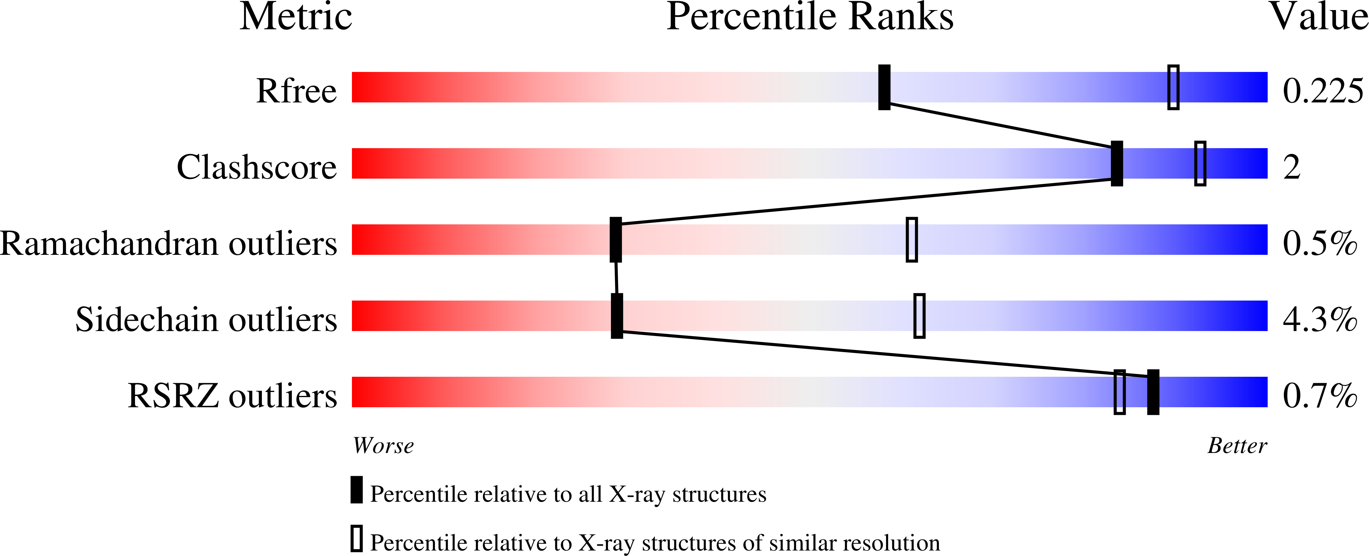
  • Method: X-RAY DIFFRACTION
  • Resolution: 2.80 Å
  • R-Value Free: 0.289 
  • R-Value Work: 0.235 
  • R-Value Observed: 0.275 

Starting Model: experimental
View more details

wwPDB Validation   3D Report Full Report


Ligand Structure Quality Assessment 


This is version 1.4 of the entry. See complete history


Literature

Structural insight into the altered substrate specificity of human cytochrome P450 2A6 mutants.

Sansen, S.Hsu, M.H.Stout, C.D.Johnson, E.F.

(2007) Arch Biochem Biophys 464: 197-206

  • DOI: https://doi.org/10.1016/j.abb.2007.04.028
  • Primary Citation of Related Structures:  
    2PG5, 2PG6, 2PG7

  • PubMed Abstract: 

    Human P450 2A6 displays a small active site that is well adapted for the oxidation of small planar substrates. Mutagenesis of CYP2A6 resulted in an increased catalytic efficiency for indole biotransformation to pigments and conferred a capacity to oxidize substituted indoles (Wu, Z.-L., Podust, L.M., Guengerich, F.P. J. Biol. Chem. 49 (2005) 41090-41100.). Here, we describe the structural basis that underlies the altered metabolic profile of three mutant enzymes, P450 2A6 N297Q, L240C/N297Q and N297Q/I300V. The Asn297 substitution abolishes a potential hydrogen bonding interaction with substrates in the active site, and replaces a structural water molecule between the helix B'-C region and helix I while maintaining structural hydrogen bonding interactions. The structures of the P450 2A6 N297Q/L240C and N297Q/I300V mutants provide clues as to how the protein can adapt to fit the larger substituted indoles in the active site, and enable a comparison with other P450 family 2 enzymes for which the residue at the equivalent position was seen to function in isozyme specificity, structural integrity and protein flexibility.


  • Organizational Affiliation

    Department of Molecular and Experimental Medicine, The Scripps Research Institute, 10550 N. Torrey Pines Road, MEM-255, La Jolla, CA 92037, USA.


Macromolecules
Find similar proteins by:  (by identity cutoff)  |  3D Structure
Entity ID: 1
MoleculeChains Sequence LengthOrganismDetailsImage
Cytochrome P450 2A6
A, B, C, D
476Homo sapiensMutation(s): 3 
Gene Names: CYP2A6
EC: 1.14.14.1
UniProt & NIH Common Fund Data Resources
Find proteins for Q16696 (Homo sapiens)
Explore Q16696 
Go to UniProtKB:  Q16696
PHAROS:  Q16696
GTEx:  ENSG00000197838 
Entity Groups  
Sequence Clusters30% Identity50% Identity70% Identity90% Identity95% Identity100% Identity
UniProt GroupQ16696
Sequence Annotations
Expand
  • Reference Sequence
Experimental Data & Validation

Experimental Data

  • Method: X-RAY DIFFRACTION
  • Resolution: 2.80 Å
  • R-Value Free: 0.289 
  • R-Value Work: 0.235 
  • R-Value Observed: 0.275 
  • Space Group: P 1 21 1
Unit Cell:
Length ( Å )Angle ( ˚ )
a = 70.889α = 90
b = 159.389β = 91.92
c = 104.101γ = 90
Software Package:
Software NamePurpose
SCALAdata processing
CNSrefinement
PDB_EXTRACTdata extraction
Blu-Icedata collection
MOSFLMdata reduction
SCALAdata scaling
CNSphasing

Structure Validation

View Full Validation Report


Ligand Structure Quality Assessment 


Entry History 

Deposition Data

Revision History  (Full details and data files)

  • Version 1.0: 2007-07-24
    Type: Initial release
  • Version 1.1: 2008-05-01
    Changes: Version format compliance
  • Version 1.2: 2011-07-13
    Changes: Version format compliance
  • Version 1.3: 2021-10-20
    Changes: Database references, Derived calculations
  • Version 1.4: 2023-08-30
    Changes: Data collection, Refinement description