2PRF | pdb_00002prf

THREE DIMENSIONAL SOLUTION STRUCTURE OF ACANTHAMOEBA PROFILIN I


Experimental Data Snapshot

  • Method: SOLUTION NMR
  • Conformers Submitted: 19 

wwPDB Validation 3D Report Full Report

Validation slider image for 2PRF

This is version 1.4 of the entry. See complete history

Literature

Three-dimensional solution structure of Acanthamoeba profilin-I.

Vinson, V.K.Archer, S.J.Lattman, E.E.Pollard, T.D.Torchia, D.A.

(1993) J Cell Biol 122: 1277-1283

  • DOI: https://doi.org/10.1083/jcb.122.6.1277
  • Primary Citation Related Structures: 
    2PRF

  • PubMed Abstract: 

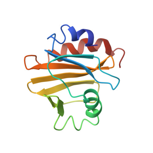
    We have determined a medium resolution three-dimensional solution structure of Acanthamoeba profilin-I by multidimensional nuclear magnetic resonance spectroscopy. This 13-kD actin binding protein consists of a five stranded antiparallel beta sheet flanked by NH2- and COOH-terminal helices on one face and by a third helix and a two stranded beta sheet on the other face. Data from actin-profilin cross-linking experiments and the localization of conserved residues between profilins in different phyla indicate that actin binding occurs on the molecular face occupied by the terminal helices. The other face of the molecule contains the residues that differ between Acanthamoeba profilins-I and II and may be important in determining the difference in polyphosphoinositide binding between these isoforms. This suggests that lipids and actin bind to different faces of the molecule.


  • Organizational Affiliation
    • Department of Cell Biology and Anatomy, Johns Hopkins University School of Medicine, Baltimore, Maryland 21205.

Macromolecule Content 

  • Total Structure Weight: 12.96 kDa 
  • Atom Count: 916 
  • Modeled Residue Count: 125 
  • Deposited Residue Count: 125 
  • Unique protein chains: 1

Macromolecules

Find similar proteins by:|  3D Structure
Entity ID: 1
MoleculeChains  Sequence LengthOrganismDetailsImage
PROFILIN IA125Acanthamoeba sp.Mutation(s): 0 
UniProt
Find proteins for P68696 (Acanthamoeba castellanii)
Explore P68696 
Go to UniProtKB:  P68696
Entity Groups
Sequence Clusters30% Identity50% Identity70% Identity90% Identity95% Identity100% Identity
UniProt GroupP68696
Sequence Annotations
Expand
Reference Sequence

Experimental Data & Validation

Experimental Data

  • Method: SOLUTION NMR
  • Conformers Submitted: 19 

Structure Validation

View Full Validation Report


Entry History 

Deposition Data

Revision History  (Full details and data files)

  • Version 1.0: 1994-05-31
    Type: Initial release
  • Version 1.1: 2008-03-25
    Changes: Version format compliance
  • Version 1.2: 2011-07-13
    Changes: Version format compliance
  • Version 1.3: 2017-11-29
    Changes: Derived calculations, Other
  • Version 1.4: 2024-05-01
    Changes: Data collection, Database references