2PSY

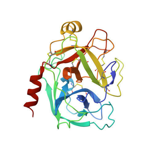
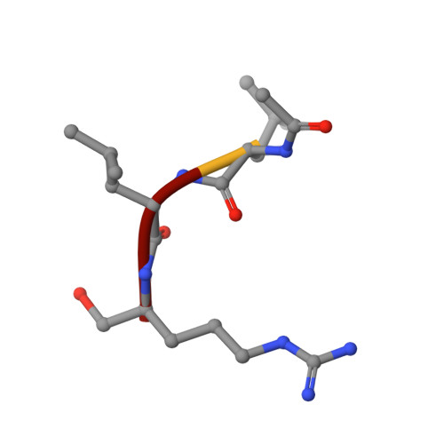
Crystal Structure of Human Kallikrein 5 in complex with Leupeptin and Zinc


Experimental Data Snapshot

  • Method: X-RAY DIFFRACTION
  • Resolution: 2.30 Å
  • R-Value Free: 0.235 
  • R-Value Work: 0.210 
  • R-Value Observed: 0.210 

Starting Model: experimental
View more details

wwPDB Validation   3D Report Full Report


This is version 2.2 of the entry. See complete history


Literature

Structural basis of the zinc inhibition of human tissue kallikrein 5.

Debela, M.Goettig, P.Magdolen, V.Huber, R.Schechter, N.M.Bode, W.

(2007) J Mol Biol 373: 1017-1031

  • DOI: https://doi.org/10.1016/j.jmb.2007.08.042
  • Primary Citation of Related Structures:  
    2PSX, 2PSY

  • PubMed Abstract: 

    Human kallikrein 5 (hK5) is a member of the tissue kallikrein family of serine peptidases. It has trypsin-like substrate specificity, is inhibited by metal ions, and is abundantly expressed in human skin, where it is believed to play a central role in desquamation. To further understand the interaction of hK5 with substrates and metal ions, active recombinant hK5 was crystallized in complex with the tripeptidyl aldehyde inhibitor leupeptin, and structures at 2.3 A resolution were obtained with and without Zn2+. While the overall structure and the specificity of S1 pocket for basic side-chains were similar to that of hK4, a closely related family member, both differed in their interaction with Zn2+. Unlike hK4, the 75-loop of hK5 is not structured to bind a Zn2+. Instead, Zn2+ binds adjacent to the active site, becoming coordinated by the imidazole rings of His99 and His96 not present in hK4. This zinc binding is accompanied by a large shift in the backbone conformation of the 99-loop and by large movements of both His side-chains. Modeling studies show that in the absence of bound leupeptin, Zn2+ is likely further coordinated by the imidazolyl side-chain of the catalytic His57 which can, similar to equivalent His57 imidazole groups in the related rat kallikrein proteinase tonin and in an engineered metal-binding rat trypsin, rotate out of its triad position to provide the third co-ordination site of the bound Zn2+, rendering Zn2+-bound hK5 inactive. In solution, this mode of binding likely occurs in the presence of free and substrate saturated hK5, as kinetic analyses of Zn2+ inhibition indicate a non-competitive mechanism. Supporting the His57 re-orientation, Zn2+ does not fully inhibit hK5 hydrolysis of tripeptidyl substrates containing a P2-His residue. The P2 and His57 imidazole groups would lie next to each other in the enzyme-substrate complex, indicating that incomplete inhibition is due to competition between both imidazole groups for Zn2+. The His96-99-57 triad is thus suggested to be responsible for the Zn2+-mediated inhibition of hK5 catalysis.


  • Organizational Affiliation

    Max-Planck-Institut für Biochemie, Proteinase Research Group, Am Klopferspitz 18, 82152 Martinsried, Germany.


Macromolecules
Find similar proteins by:  (by identity cutoff)  |  3D Structure
Entity ID: 1
MoleculeChains Sequence LengthOrganismDetailsImage
Kallikrein-5227Homo sapiensMutation(s): 0 
Gene Names: KLK5
EC: 3.4.21
UniProt & NIH Common Fund Data Resources
Find proteins for Q9Y337 (Homo sapiens)
Explore Q9Y337 
Go to UniProtKB:  Q9Y337
PHAROS:  Q9Y337
GTEx:  ENSG00000167754 
Entity Groups  
Sequence Clusters30% Identity50% Identity70% Identity90% Identity95% Identity100% Identity
UniProt GroupQ9Y337
Glycosylation
Glycosylation Sites: 1Go to GlyGen: Q9Y337-1
Sequence Annotations
Expand
  • Reference Sequence

Find similar proteins by:  Sequence   |   3D Structure  

Entity ID: 2
MoleculeChains Sequence LengthOrganismDetailsImage
LEUPEPTIN4Streptomyces roseusMutation(s): 0 
Sequence Annotations
Expand
  • Reference Sequence
Oligosaccharides

Help

Entity ID: 3
MoleculeChains Length2D Diagram Glycosylation3D Interactions
2-acetamido-2-deoxy-beta-D-glucopyranose-(1-4)-2-acetamido-2-deoxy-beta-D-glucopyranose
C
2N-Glycosylation
Glycosylation Resources
GlyTouCan:  G42666HT
GlyCosmos:  G42666HT
GlyGen:  G42666HT
Biologically Interesting Molecules (External Reference) 1 Unique
Experimental Data & Validation

Experimental Data

  • Method: X-RAY DIFFRACTION
  • Resolution: 2.30 Å
  • R-Value Free: 0.235 
  • R-Value Work: 0.210 
  • R-Value Observed: 0.210 
  • Space Group: C 1 2 1
Unit Cell:
Length ( Å )Angle ( ˚ )
a = 126.532α = 90
b = 66.543β = 95.14
c = 39.58γ = 90
Software Package:
Software NamePurpose
CNSrefinement
DENZOdata reduction
SCALEPACKdata scaling
PHASERphasing

Structure Validation

View Full Validation Report


Entry History 

Deposition Data

Revision History  (Full details and data files)

  • Version 1.0: 2007-09-11
    Type: Initial release
  • Version 1.1: 2011-07-13
    Changes: Atomic model, Database references, Derived calculations, Non-polymer description, Structure summary, Version format compliance
  • Version 1.2: 2012-12-12
    Changes: Other
  • Version 2.0: 2020-07-29
    Type: Remediation
    Reason: Carbohydrate remediation
    Changes: Advisory, Atomic model, Data collection, Derived calculations, Structure summary
  • Version 2.1: 2023-08-30
    Changes: Advisory, Data collection, Database references, Refinement description, Structure summary
  • Version 2.2: 2024-11-06
    Changes: Structure summary