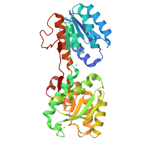
2QW1

Glucose/galactose binding protein bound to 3-O-methyl D-glucose


Experimental Data Snapshot

  • Method: X-RAY DIFFRACTION
  • Resolution: 1.70 Å
  • R-Value Free: 0.204 
  • R-Value Work: 0.180 
  • R-Value Observed: 0.182 

Starting Model: experimental
View more details

wwPDB Validation   3D Report Full Report


Ligand Structure Quality Assessment 


This is version 1.3 of the entry. See complete history


Literature

Structure-based design of a periplasmic binding protein antagonist that prevents domain closure.

Borrok, M.J.Zhu, Y.Forest, K.T.Kiessling, L.L.

(2009) ACS Chem Biol 4: 447-456

  • DOI: https://doi.org/10.1021/cb900021q
  • Primary Citation of Related Structures:  
    2QW1

  • PubMed Abstract: 

    Many receptors undergo ligand-induced conformational changes to initiate signal transduction. Periplasmic binding proteins (PBPs) are bacterial receptors that exhibit dramatic conformational changes upon ligand binding. These proteins mediate a wide variety of fundamental processes including transport, chemotaxis, and quorum sensing. Despite the importance of these receptors, no PBP antagonists have been identified and characterized. In this study, we identify 3-O-methyl-d-glucose as an antagonist of glucose/galactose-binding protein and demonstrate that it inhibits glucose chemotaxis in E. coli. Using small-angle X-ray scattering and X-ray crystallography, we show that this antagonist acts as a wedge. It prevents the large-scale domain closure that gives rise to the active signaling state. Guided by these results and the structures of open and closed glucose/galactose-binding protein, we designed and synthesized an antagonist composed of two linked glucose residues. These findings provide a blueprint for the design of new bacterial PBP inhibitors. Given the key role of PBPs in microbial physiology, we anticipate that PBP antagonists will have widespread uses as probes and antimicrobial agents.


  • Organizational Affiliation

    Department of Biochemistry, University of Wisconsin, Madison, Wisconsin 53706, USA.


Macromolecules
Find similar proteins by:  (by identity cutoff)  |  3D Structure
Entity ID: 1
MoleculeChains Sequence LengthOrganismDetailsImage
D-galactose-binding periplasmic protein309Escherichia coliMutation(s): 0 
Gene Names: mglB
UniProt
Find proteins for P0AEE5 (Escherichia coli (strain K12))
Explore P0AEE5 
Go to UniProtKB:  P0AEE5
Entity Groups  
Sequence Clusters30% Identity50% Identity70% Identity90% Identity95% Identity100% Identity
UniProt GroupP0AEE5
Sequence Annotations
Expand
  • Reference Sequence
Experimental Data & Validation

Experimental Data

  • Method: X-RAY DIFFRACTION
  • Resolution: 1.70 Å
  • R-Value Free: 0.204 
  • R-Value Work: 0.180 
  • R-Value Observed: 0.182 
  • Space Group: P 21 21 21
Unit Cell:
Length ( Å )Angle ( ˚ )
a = 56.93α = 90
b = 74.66β = 90
c = 110.07γ = 90
Software Package:
Software NamePurpose
REFMACrefinement
PROTEUM PLUSdata collection
SAINTdata reduction
PROTEUM PLUSdata scaling
CNSphasing

Structure Validation

View Full Validation Report


Ligand Structure Quality Assessment 


Entry History 

Deposition Data

Revision History  (Full details and data files)

  • Version 1.0: 2008-08-05
    Type: Initial release
  • Version 1.1: 2011-07-13
    Changes: Version format compliance
  • Version 1.2: 2020-07-29
    Type: Remediation
    Reason: Carbohydrate remediation
    Changes: Data collection, Derived calculations
  • Version 1.3: 2023-08-30
    Changes: Data collection, Database references, Refinement description, Structure summary