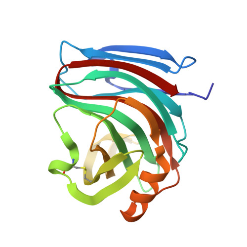
2QZ2

Crystal structure of a glycoside hydrolase family 11 xylanase from Aspergillus niger in complex with xylopentaose


Experimental Data Snapshot

  • Method: X-RAY DIFFRACTION
  • Resolution: 2.80 Å
  • R-Value Free: 0.277 
  • R-Value Work: 0.221 
  • R-Value Observed: 0.226 

Starting Model: experimental
View more details

wwPDB Validation   3D Report Full Report


This is version 2.3 of the entry. See complete history


Literature

Crystallographic analysis shows substrate binding at the -3 to +1 active-site subsites and at the surface of glycoside hydrolase family 11 endo-1,4-beta-xylanases.

Vandermarliere, E.Bourgois, T.M.Rombouts, S.Van Campenhout, S.Volckaert, G.Strelkov, S.V.Delcour, J.A.Rabijns, A.Courtin, C.M.

(2008) Biochem J 410: 71-79

  • DOI: https://doi.org/10.1042/BJ20071128
  • Primary Citation of Related Structures:  
    2QZ2, 2QZ3, 2Z79

  • PubMed Abstract: 

    GH 11 (glycoside hydrolase family 11) xylanases are predominant enzymes in the hydrolysis of heteroxylan, an abundant structural polysaccharide in the plant cell wall. To gain more insight into the protein-ligand interactions of the glycone as well as the aglycone subsites of these enzymes, catalytically incompetent mutants of the Bacillus subtilis and Aspergillus niger xylanases were crystallized, soaked with xylo-oligosaccharides and subjected to X-ray analysis. For both xylanases, there was clear density for xylose residues in the -1 and -2 subsites. In addition, for the B. subtilis xylanase, there was also density for xylose residues in the -3 and +1 subsite showing the spanning of the -1/+1 subsites. These results, together with the observation that some residues in the aglycone subsites clearly adopt a different conformation upon substrate binding, allowed us to identify the residues important for substrate binding in the aglycone subsites. In addition to substrate binding in the active site of the enzymes, the existence of an unproductive second ligand-binding site located on the surface of both the B. subtilis and A. niger xylanases was observed. This extra binding site may have a function similar to the separate carbohydrate-binding modules of other glycoside hydrolase families.


  • Organizational Affiliation

    Laboratory for Biocrystallography, Department of Pharmaceutical Sciences, Katholieke Universiteit Leuven, Herestraat 49, O&N II, bus 822, 3000 Leuven, Belgium. Elien.Vandermarliere@pharm.kuleuven.be


Macromolecules
Find similar proteins by:  (by identity cutoff)  |  3D Structure
Entity ID: 1
MoleculeChains Sequence LengthOrganismDetailsImage
Endo-1,4-beta-xylanase I184Aspergillus nigerMutation(s): 1 
EC: 3.2.1.8
UniProt
Find proteins for P55329 (Aspergillus niger)
Explore P55329 
Go to UniProtKB:  P55329
Entity Groups  
Sequence Clusters30% Identity50% Identity70% Identity90% Identity95% Identity100% Identity
UniProt GroupP55329
Sequence Annotations
Expand
  • Reference Sequence
Oligosaccharides

Help

Entity ID: 2
MoleculeChains Length2D Diagram Glycosylation3D Interactions
beta-D-xylopyranose-(1-4)-beta-D-xylopyranose
B
2N/A
Glycosylation Resources
GlyTouCan:  G87728WL
GlyCosmos:  G87728WL
Entity ID: 3
MoleculeChains Length2D Diagram Glycosylation3D Interactions
beta-D-xylopyranose-(1-4)-beta-D-xylopyranose-(1-4)-beta-D-xylopyranose-(1-4)-beta-D-xylopyranose
C
4N/A
Glycosylation Resources
GlyTouCan:  G60650FG
GlyCosmos:  G60650FG
Small Molecules
Ligands 1 Unique
IDChains Name / Formula / InChI Key2D Diagram3D Interactions
NA
Query on NA

Download Ideal Coordinates CCD File 
D [auth A]SODIUM ION
Na
FKNQFGJONOIPTF-UHFFFAOYSA-N
Biologically Interesting Molecules (External Reference) 1 Unique
Experimental Data & Validation

Experimental Data

  • Method: X-RAY DIFFRACTION
  • Resolution: 2.80 Å
  • R-Value Free: 0.277 
  • R-Value Work: 0.221 
  • R-Value Observed: 0.226 
  • Space Group: P 65 2 2
Unit Cell:
Length ( Å )Angle ( ˚ )
a = 67.326α = 90
b = 67.326β = 90
c = 165.401γ = 120
Software Package:
Software NamePurpose
REFMACrefinement
MAR345dtbdata collection
DENZOdata reduction
SCALEPACKdata scaling
PHASERphasing

Structure Validation

View Full Validation Report


Entry History 

Deposition Data

Revision History  (Full details and data files)

  • Version 1.0: 2007-12-25
    Type: Initial release
  • Version 1.1: 2011-07-13
    Changes: Version format compliance
  • Version 2.0: 2020-07-29
    Type: Remediation
    Reason: Carbohydrate remediation
    Changes: Advisory, Atomic model, Data collection, Derived calculations, Structure summary
  • Version 2.1: 2021-11-10
    Changes: Database references, Derived calculations, Structure summary
  • Version 2.2: 2023-10-25
    Changes: Data collection, Refinement description
  • Version 2.3: 2024-10-09
    Changes: Structure summary