2V26 | pdb_00002v26

Myosin VI (MD) pre-powerstroke state (Mg.ADP.VO4)


Experimental Data Snapshot

  • Method: X-RAY DIFFRACTION
  • Resolution: 1.75 Å
  • R-Value Free: 
    0.227 (Depositor) 
  • R-Value Work: 
    0.202 (Depositor) 
  • R-Value Observed: 
    0.205 (Depositor) 

Starting Model: experimental
View more details

wwPDB Validation 3D Report Full Report

Validation slider image for 2V26

Ligand Structure Quality Assessment 


This is version 1.3 of the entry. See complete history

Literature

The Structural Basis for the Large Powerstroke of Myosin Vi.

Menetrey, J.Llinas, P.Mukherjea, M.Sweeney, H.L.Houdusse, A.

(2007) Cell 131: 300

  • DOI: https://doi.org/10.1016/j.cell.2007.08.027
  • Primary Citation Related Structures: 
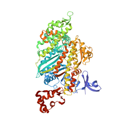
    2V26

  • PubMed Abstract: 

    Due to a unique addition to the lever arm-positioning region (converter), class VI myosins move in the opposite direction (toward the minus-end of actin filaments) compared to other characterized myosin classes. However, the large size of the myosin VI lever arm swing (powerstroke) cannot be explained by our current view of the structural transitions that occur within the myosin motor. We have solved the crystal structure of a fragment of the myosin VI motor in the structural state that represents the starting point for movement on actin; the pre-powerstroke state. Unexpectedly, the converter itself rearranges to achieve a conformation that has not been seen for other myosins. This results in a much larger powerstroke than is achievable without the converter rearrangement. Moreover, it provides a new mechanism that could be exploited to increase the powerstroke of yet to be characterized plus-end-directed myosin classes.


  • Organizational Affiliation
    • Structural Motility, Institut Curie CNRS, UMR144, 26 rue d'Ulm, 75248 Paris cedex 05, France.

Macromolecule Content 

  • Total Structure Weight: 90.63 kDa 
  • Atom Count: 6,665 
  • Modeled Residue Count: 747 
  • Deposited Residue Count: 784 
  • Unique protein chains: 1

Macromolecules

Find similar proteins by:|  3D Structure
Entity ID: 1
MoleculeChains  Sequence LengthOrganismDetailsImage
MYOSIN VI784Sus scrofaMutation(s): 0 
UniProt
Find proteins for Q29122 (Sus scrofa)
Explore Q29122 
Go to UniProtKB:  Q29122
Entity Groups
Sequence Clusters30% Identity50% Identity70% Identity90% Identity95% Identity100% Identity
UniProt GroupQ29122
Sequence Annotations
Expand
Reference Sequence

Small Molecules

Ligands 5 Unique
IDChains Name / Formula / InChI Key2D Diagram3D Interactions
ADP

Query on ADP


Download:Ideal Coordinates CCD File
B [auth A]ADENOSINE-5'-DIPHOSPHATE
C10 H15 N5 O10 P2
XTWYTFMLZFPYCI-KQYNXXCUSA-N
VO4

Query on VO4


Download:Ideal Coordinates CCD File
C [auth A]VANADATE ION
O4 V
LSGOVYNHVSXFFJ-UHFFFAOYSA-N
SO4

Query on SO4


Download:Ideal Coordinates CCD File
K [auth A],
L [auth A]
SULFATE ION
O4 S
QAOWNCQODCNURD-UHFFFAOYSA-L
EDO

Query on EDO


Download:Ideal Coordinates CCD File
D [auth A]
E [auth A]
F [auth A]
G [auth A]
H [auth A]
D [auth A],
E [auth A],
F [auth A],
G [auth A],
H [auth A],
I [auth A],
J [auth A]
1,2-ETHANEDIOL
C2 H6 O2
LYCAIKOWRPUZTN-UHFFFAOYSA-N
MG

Query on MG


Download:Ideal Coordinates CCD File
M [auth A]MAGNESIUM ION
Mg
JLVVSXFLKOJNIY-UHFFFAOYSA-N

Experimental Data & Validation

Experimental Data

  • Method: X-RAY DIFFRACTION
  • Resolution: 1.75 Å
  • R-Value Free:  0.227 (Depositor) 
  • R-Value Work:  0.202 (Depositor) 
  • R-Value Observed: 0.205 (Depositor) 
Space Group: P 21 21 21
Unit Cell:
Length ( Å )Angle ( ˚ )
a = 93.512α = 90
b = 93.845β = 90
c = 100.976γ = 90
Software Package:
Software NamePurpose
REFMACrefinement
MOSFLMdata reduction
SCALAdata scaling
AMoREphasing

Structure Validation

View Full Validation Report


Ligand Structure Quality Assessment 


Entry History 

Deposition Data

Revision History  (Full details and data files)

  • Version 1.0: 2007-11-20
    Type: Initial release
  • Version 1.1: 2011-05-08
    Changes: Version format compliance
  • Version 1.2: 2011-07-13
    Changes: Version format compliance
  • Version 1.3: 2023-12-13
    Changes: Data collection, Database references, Derived calculations, Other, Refinement description