2VQX | pdb_00002vqx

Precursor of Protealysin, Metalloproteinase from Serratia proteamaculans.


Experimental Data Snapshot

  • Method: X-RAY DIFFRACTION
  • Resolution: 1.82 Å
  • R-Value Free: 
    0.202 (Depositor), 0.208 (DCC) 
  • R-Value Work: 
    0.170 (Depositor), 0.176 (DCC) 
  • R-Value Observed: 
    0.171 (Depositor) 

Starting Model: experimental
View more details

wwPDB Validation 3D Report Full Report

Validation slider image for 2VQX

This is version 1.4 of the entry. See complete history

Literature

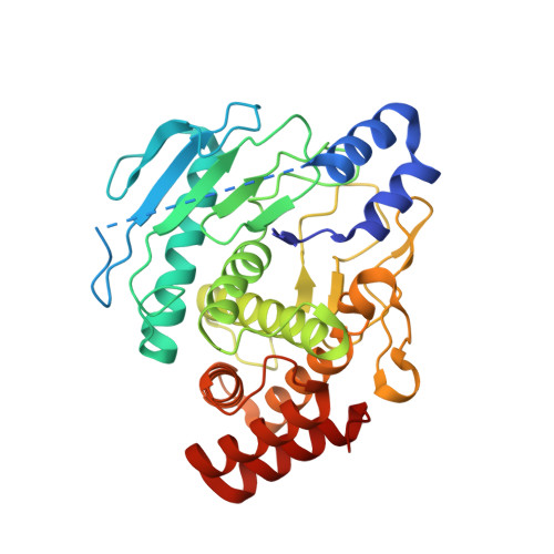
Crystal Structure of the Protealysin Precursor: Insights Into Propeptide Function.

Demidyuk, I.V.Gromova, T.Y.Polyakov, K.M.Melik-Adamyan, W.R.Kuranova, I.P.Kostrov, S.V.

(2010) J Biological Chem 285: 2003

  • DOI: https://doi.org/10.1074/jbc.M109.015396
  • Primary Citation Related Structures: 
    2VQX

  • PubMed Abstract: 

    Protealysin (PLN) belongs to the M4 family of peptidases that are commonly known as thermolysin-like proteases (TLPs). All TLPs are synthesized as precursors containing N-terminal propeptides. According to the primary structure of the N-terminal propeptides, the family is divided into two distinct groups. Representatives of the first group including thermolysin and all TLPs with known three-dimensional structures have long prosequences ( approximately 200 amino acids). Enzymes of the second group, whose prototype is protealysin, have short ( approximately 50 amino acids) propeptides. Here, we present the 1.8 A crystal structure of PLN precursor (proPLN), which is the first three-dimensional structure of a TLP precursor. Whereas the structure of the catalytic domain of proPLN is similar overall to previously reported structures of mature TLPs, it has specific features, including the absence of calcium-binding sites, and different structures of the N-terminal region and substrate-binding site. PLN propeptide forms a separate domain in the precursor and likely acts as an inhibitor that blocks the substrate-binding site and fixes the "open" conformation of the active site, which is unfavorable for catalysis. Furthermore the conserved PPL motif identified in our previous studies directly interacts with the S' subsites of the active center being a critical element of the propeptide-catalytic domain interface. Comparison of the primary structures of TLPs with short propeptides suggests that the specific features revealed in the proPLN crystal structure are typical for all protealysin-like enzymes. Thus, such proteins can be considered as a separate subfamily of TLPs.


  • Organizational Affiliation
    • Institute of Molecular Genetics, Russian Academy of Sciences, Kurchatov Sq. 2, Moscow 123182, Russia. duk@img.ras.ru

Macromolecule Content 

  • Total Structure Weight: 37.52 kDa 
  • Atom Count: 2,805 
  • Modeled Residue Count: 322 
  • Deposited Residue Count: 341 
  • Unique protein chains: 1

Macromolecules

Find similar proteins by:|  3D Structure
Entity ID: 1
MoleculeChains  Sequence LengthOrganismDetailsImage
METALLOPROTEINASE341Serratia proteamaculansMutation(s): 1 
EC: 3.4.24
UniProt
Find proteins for Q5MJ80 (Serratia proteamaculans)
Explore Q5MJ80 
Go to UniProtKB:  Q5MJ80
Entity Groups
Sequence Clusters30% Identity50% Identity70% Identity90% Identity95% Identity100% Identity
UniProt GroupQ5MJ80
Sequence Annotations
Expand
Reference Sequence

Small Molecules

Ligands 1 Unique
IDChains Name / Formula / InChI Key2D Diagram3D Interactions
ZN

Query on ZN


Download:Ideal Coordinates CCD File
B [auth A]ZINC ION
Zn
PTFCDOFLOPIGGS-UHFFFAOYSA-N

Experimental Data & Validation

Experimental Data

  • Method: X-RAY DIFFRACTION
  • Resolution: 1.82 Å
  • R-Value Free:  0.202 (Depositor), 0.208 (DCC) 
  • R-Value Work:  0.170 (Depositor), 0.176 (DCC) 
  • R-Value Observed: 0.171 (Depositor) 
Space Group: P 21 21 2
Unit Cell:
Length ( Å )Angle ( ˚ )
a = 70.76α = 90
b = 78.65β = 90
c = 59.28γ = 90
Software Package:
Software NamePurpose
XDSdata reduction
XDSdata scaling
BALBESphasing
MOLREPphasing
REFMACrefinement

Structure Validation

View Full Validation Report


Entry History 

Deposition Data

Revision History  (Full details and data files)

  • Version 1.0: 2009-05-26
    Type: Initial release
  • Version 1.1: 2011-05-08
    Changes: Version format compliance
  • Version 1.2: 2011-07-13
    Changes: Version format compliance
  • Version 1.3: 2019-05-15
    Changes: Data collection, Experimental preparation, Other
  • Version 1.4: 2023-12-13
    Changes: Data collection, Database references, Derived calculations, Other, Refinement description