


Crystal Structure of Cbpf in Complex with Atropine by Soaking
Silva-Martin, N., Hermoso, J.A., Sanz, J.M.To be published.
Experimental Data Snapshot
Starting Model: experimental
View more details
Entity ID: 1 | |||||
|---|---|---|---|---|---|
| Molecule | Chains | Sequence Length | Organism | Details | Image |
| CHOLINE-BINDING PROTEIN F | 311 | Streptococcus pneumoniae R6 | Mutation(s): 0 | 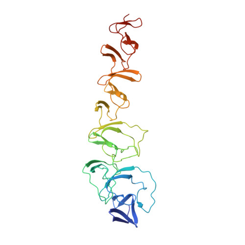 | |
UniProt | |||||
Entity Groups | |||||
| Sequence Clusters | 30% Identity50% Identity70% Identity90% Identity95% Identity100% Identity | ||||
| UniProt Group | Q8DR52 | ||||
Sequence AnnotationsExpand | |||||
Reference Sequence | |||||
| Ligands 4 Unique | |||||
|---|---|---|---|---|---|
| ID | Chains | Name / Formula / InChI Key | 2D Diagram | 3D Interactions | |
| OIN Download:Ideal Coordinates CCD File | D [auth A], G [auth A] | (1R,5S)-8-METHYL-8-AZABICYCLO[3.2.1]OCT-3-YL (2R)-3-HYDROXY-2-PHENYLPROPANOATE C17 H23 N O3 RKUNBYITZUJHSG-QKPAOTATSA-N |  | ||
| CHT Download:Ideal Coordinates CCD File | B [auth A] H [auth A] I [auth A] L [auth A] M [auth A] | CHOLINE ION C5 H14 N O OEYIOHPDSNJKLS-UHFFFAOYSA-N |  | ||
| SO4 Download:Ideal Coordinates CCD File | C [auth A] | SULFATE ION O4 S QAOWNCQODCNURD-UHFFFAOYSA-L |  | ||
| GOL Download:Ideal Coordinates CCD File | E [auth A], F [auth A], J [auth A], K [auth A] | GLYCEROL C3 H8 O3 PEDCQBHIVMGVHV-UHFFFAOYSA-N |  | ||
| Length ( Å ) | Angle ( ˚ ) |
|---|---|
| a = 52.409 | α = 90 |
| b = 116.752 | β = 90 |
| c = 73.889 | γ = 90 |
| Software Name | Purpose |
|---|---|
| CNS | refinement |
| XDS | data reduction |
| SCALEPACK | data scaling |
| MOLREP | phasing |


