2XA7 | pdb_00002xa7

AP2 clathrin adaptor core in active complex with cargo peptides


Experimental Data Snapshot

  • Method: X-RAY DIFFRACTION
  • Resolution: 3.10 Å
  • R-Value Free: 
    0.285 (Depositor), 0.282 (DCC) 
  • R-Value Work: 
    0.236 (Depositor) 
  • R-Value Observed: 
    0.239 (Depositor) 

wwPDB Validation 3D Report Full Report

Validation slider image for 2XA7

This is version 1.3 of the entry. See complete history. 

Literature

A Large Scale Conformational Change Couples Membrane Recruitment to Cargo Binding in the Ap2 Clathrin Adaptor Complex

Jackson, L.P., Kelly, B.T., Mccoy, A.J., Gaffry, T., James, L.C., Collins, B.M., Honing, S., Evans, P.R., Owen, D.J.

(2010) Cell 141: 1241

  • DOI: https://doi.org/10.1016/j.cell.2010.05.006
  • Primary Citation Related Structures: 
    2XA7

  • PubMed Abstract: 

    The AP2 adaptor complex (alpha, beta2, sigma2, and mu2 subunits) crosslinks the endocytic clathrin scaffold to PtdIns4,5P(2)-containing membranes and transmembrane protein cargo. In the "locked" cytosolic form, AP2's binding sites for the two endocytic motifs, YxxPhi on the C-terminal domain of mu2 (C-mu2) and [ED]xxxL[LI] on sigma2, are blocked by parts of beta2. Using protein crystallography, we show that AP2 undergoes a large conformational change in which C-mu2 relocates to an orthogonal face of the complex, simultaneously unblocking both cargo-binding sites; the previously unstructured mu2 linker becomes helical and binds back onto the complex. This structural rearrangement results in AP2's four PtdIns4,5P(2)- and two endocytic motif-binding sites becoming coplanar, facilitating their simultaneous interaction with PtdIns4,5P(2)/cargo-containing membranes. Using a range of biophysical techniques, we show that the endocytic cargo binding of AP2 is driven by its interaction with PtdIns4,5P(2)-containing membranes.


  • Organizational Affiliation: 
    • Cambridge Institute for Medical Research, Department of Clinical Biochemistry, University of Cambridge, Hills Road, Cambridge CB2 0XY, UK.

Macromolecule Content 

  • Total Structure Weight: 206.12 kDa 
  • Atom Count: 14,137 
  • Modeled Residue Count: 1,768 
  • Deposited Residue Count: 1,807 
  • Unique protein chains: 5

Macromolecules

Find similar proteins by:|  3D Structure
Entity ID: 1
MoleculeChains  Sequence LengthOrganismDetailsImage
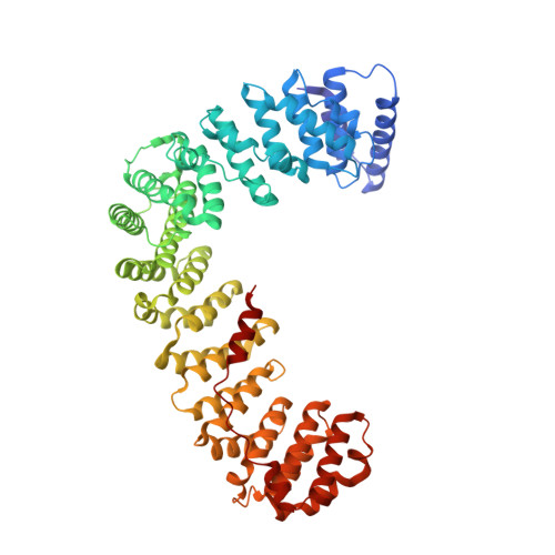
ADAPTOR-RELATED PROTEIN COMPLEX 2, ALPHA 2 SUBUNIT621Rattus norvegicusMutation(s): 0 
UniProt
Find proteins for P18484 (Rattus norvegicus)
Explore P18484 
Go to UniProtKB:  P18484
Entity Groups
Sequence Clusters30% Identity50% Identity70% Identity90% Identity95% Identity100% Identity
UniProt GroupP18484
Sequence Annotations
Expand
Reference Sequence
Find similar proteins by:|  3D Structure
Entity ID: 2
MoleculeChains  Sequence LengthOrganismDetailsImage
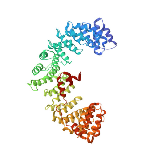
AP-2 COMPLEX SUBUNIT BETA592Homo sapiensMutation(s): 0 
UniProt & NIH Common Fund Data Resources
Find proteins for P63010 (Homo sapiens)
Explore P63010 
Go to UniProtKB:  P63010
PHAROS:  P63010
GTEx:  ENSG00000006125 
Entity Groups
Sequence Clusters30% Identity50% Identity70% Identity90% Identity95% Identity100% Identity
UniProt GroupP63010
Sequence Annotations
Expand
Reference Sequence
Find similar proteins by:|  3D Structure
Entity ID: 3
MoleculeChains  Sequence LengthOrganismDetailsImage
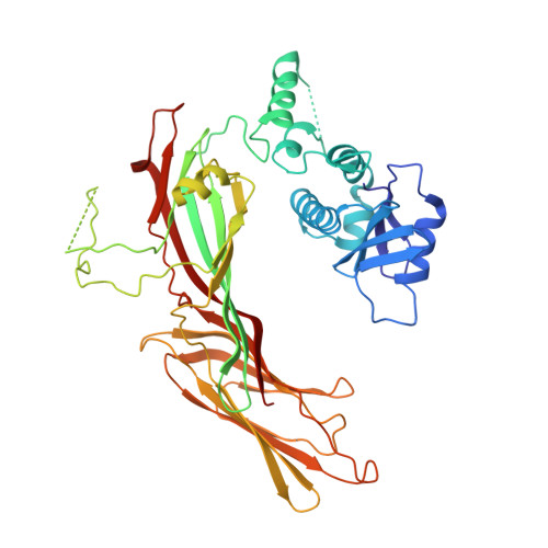
AP-2 COMPLEX SUBUNIT MU,C [auth M]446Rattus norvegicusMutation(s): 0 
UniProt
Find proteins for P84092 (Rattus norvegicus)
Explore P84092 
Go to UniProtKB:  P84092
Entity Groups
Sequence Clusters30% Identity50% Identity70% Identity90% Identity95% Identity100% Identity
UniProt GroupP84092
Sequence Annotations
Expand
Reference Sequence
Find similar proteins by:  Sequence   |   3D Structure  
Entity ID: 4
MoleculeChains  Sequence LengthOrganismDetailsImage
TGN38 CARGO PEPTIDED [auth P]6Homo sapiensMutation(s): 0 
Sequence Annotations
Expand
Reference Sequence
Find similar proteins by:|  3D Structure
Entity ID: 5
MoleculeChains  Sequence LengthOrganismDetailsImage
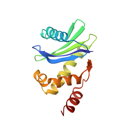
AP-2 COMPLEX SUBUNIT SIGMAE [auth S]142Mus musculusMutation(s): 0 
UniProt & NIH Common Fund Data Resources
Find proteins for P62743 (Mus musculus)
Explore P62743 
Go to UniProtKB:  P62743
IMPC:  MGI:2141861
Entity Groups
Sequence Clusters30% Identity50% Identity70% Identity90% Identity95% Identity100% Identity
UniProt GroupP62743
Sequence Annotations
Expand
Reference Sequence

Experimental Data & Validation

Experimental Data

  • Method: X-RAY DIFFRACTION
  • Resolution: 3.10 Å
  • R-Value Free:  0.285 (Depositor), 0.282 (DCC) 
  • R-Value Work:  0.236 (Depositor) 
  • R-Value Observed: 0.239 (Depositor) 
Space Group: H 3
Unit Cell:
Length ( Å )Angle ( ˚ )
a = 255.316α = 90
b = 255.316β = 90
c = 156.754γ = 120
Software Package:
Software NamePurpose
REFMACrefinement
MOSFLMdata reduction
SCALAdata scaling
SHARPphasing

Structure Validation

View Full Validation Report


Entry History 

Deposition Data

Revision History  (Full details and data files)

  • Version 1.0: 2010-07-21
    Type: Initial release
  • Version 1.1: 2011-05-08
    Changes: Version format compliance
  • Version 1.2: 2011-07-13
    Changes: Version format compliance
  • Version 1.3: 2024-05-08
    Changes: Data collection, Database references, Derived calculations, Other