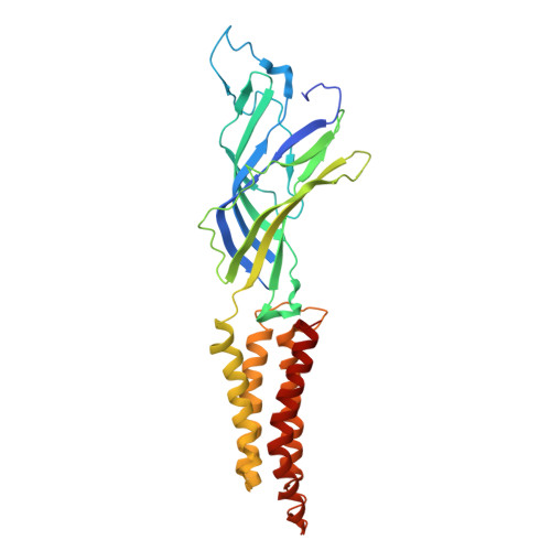
2XQ7

Pentameric ligand gated ion channel GLIC in complex with cadmium ion (Cd2+)


Experimental Data Snapshot

  • Method: X-RAY DIFFRACTION
  • Resolution: 3.40 Å
  • R-Value Free: 0.266 
  • R-Value Work: 0.242 
  • R-Value Observed: 0.243 

wwPDB Validation   3D Report Full Report


This is version 1.5 of the entry. See complete history


Literature

Structural Basis of Open Channel Block in a Prokaryotic Pentameric Ligand-Gated Ion Channel

Hilf, R.J.C.Bertozzi, C.Zimmermann, I.Reiter, A.Trauner, D.Dutzler, R.

(2010) Nat Struct Mol Biol 17: 1330

  • DOI: https://doi.org/10.1038/nsmb.1933
  • Primary Citation of Related Structures:  
    2XQ3, 2XQ4, 2XQ5, 2XQ6, 2XQ7, 2XQ8, 2XQ9, 2XQA

  • PubMed Abstract: 

    The flow of ions through cation-selective members of the pentameric ligand-gated ion channel family is inhibited by a structurally diverse class of molecules that bind to the transmembrane pore in the open state of the protein. To obtain insight into the mechanism of channel block, we have investigated the binding of positively charged inhibitors to the open channel of the bacterial homolog GLIC by using X-ray crystallography and electrophysiology. Our studies reveal the location of two regions for interactions, with larger blockers binding in the center of the membrane and divalent transition metal ions binding to the narrow intracellular pore entry. The results provide a structural foundation for understanding the interactions of the channel with inhibitors that is relevant for the entire family.


  • Organizational Affiliation

    Department of Biochemistry, University of Zürich, Zürich, Switzerland.


Macromolecules
Find similar proteins by:  (by identity cutoff)  |  3D Structure
Entity ID: 1
MoleculeChains Sequence LengthOrganismDetailsImage
GLR4197 PROTEIN
A, B, C, D, E
317Gloeobacter violaceus PCC 7421Mutation(s): 0 
Membrane Entity: Yes 
UniProt
Find proteins for Q7NDN8 (Gloeobacter violaceus (strain ATCC 29082 / PCC 7421))
Explore Q7NDN8 
Go to UniProtKB:  Q7NDN8
Entity Groups  
Sequence Clusters30% Identity50% Identity70% Identity90% Identity95% Identity100% Identity
UniProt GroupQ7NDN8
Sequence Annotations
Expand
  • Reference Sequence
Experimental Data & Validation

Experimental Data

  • Method: X-RAY DIFFRACTION
  • Resolution: 3.40 Å
  • R-Value Free: 0.266 
  • R-Value Work: 0.242 
  • R-Value Observed: 0.243 
  • Space Group: C 1 2 1
Unit Cell:
Length ( Å )Angle ( ˚ )
a = 183.314α = 90
b = 128.31β = 104.04
c = 164.374γ = 90
Software Package:
Software NamePurpose
PHENIXrefinement
XDSdata reduction
XDSdata scaling

Structure Validation

View Full Validation Report


Entry History 

Deposition Data

Revision History  (Full details and data files)

  • Version 1.0: 2010-11-10
    Type: Initial release
  • Version 1.1: 2011-05-19
    Changes: Version format compliance
  • Version 1.2: 2011-07-13
    Changes: Version format compliance
  • Version 1.3: 2019-01-30
    Changes: Data collection, Experimental preparation, Other
  • Version 1.4: 2019-02-06
    Changes: Data collection, Experimental preparation
  • Version 1.5: 2024-05-08
    Changes: Data collection, Database references, Derived calculations, Other