2XUF

CRYSTAL STRUCTURE OF MACHE-Y337A-TZ2PA6 ANTI COMPLEX (1 MTH)


Experimental Data Snapshot

  • Method: X-RAY DIFFRACTION
  • Resolution: 2.55 Å
  • R-Value Free: 
    0.221 (Depositor), 0.220 (DCC) 
  • R-Value Work: 
    0.191 (Depositor), 0.190 (DCC) 
  • R-Value Observed: 
    0.191 (Depositor) 

Starting Model: experimental
View more details

wwPDB Validation   3D Report Full Report


Ligand Structure Quality Assessment 

Created with Raphaël 2.3.0Worse 01 BetterLigand structure goodness of fit to experimental dataBest fitted TZ4Click on this verticalbar to view detailsBest fitted NAGClick on this verticalbar to view details

This is version 1.6 of the entry. See complete history


Literature

Conformational Remodeling of Femtomolar Inhibitor-Acetylcholinesterase Complexes in the Crystalline State

Bourne, Y.Radic, Z.Taylor, P.Marchot, P.

(2010) J Am Chem Soc 132: 18292

  • DOI: https://doi.org/10.1021/ja106820e
  • Primary Citation of Related Structures:  
    2XUD, 2XUF, 2XUG, 2XUH, 2XUI, 2XUJ, 2XUK, 2XUO, 2XUP, 2XUQ

  • PubMed Abstract: 

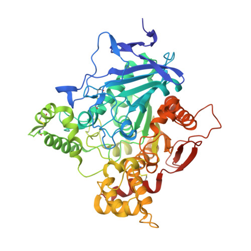
    The active center of acetylcholinesterase (AChE), a target site for competitive inhibitors, resides centrosymmetric to the subunit at the base of a deep, narrow gorge lined by aromatic residues. At the gorge entry, a peripheral site encompasses overlapping binding loci for noncompetitive inhibitors, which alter substrate access to the gorge. The click-chemistry inhibitor TZ2PA6 links the active center ligand, tacrine, to the peripheral site ligand, propidium, through a biorthogonal reaction of an acetylene and an azide that forms either a syn1 or an anti1 triazole. Compared with wild-type mouse AChE, a Tyr337Ala mutant displays full catalytic activity, albeit with 2-3 orders of magnitude higher affinities for the TZ2PA6 syn1 and anti1 regioisomers, reflected in low femtomolar K(d) values, diffusion-limited association, and dissociation half-times greater than 1 month and 1 week, respectively. Three structures of each of the co-crystallized syn1 and anti1 complexes of the Tyr337Ala mutant were solved at three distinct times of crystal maturation, consistent with or exceeding the half-lives of the complexes in solution, while crystalline complexes obtained from soaked Tyr337Ala crystals led to picturing "freshly formed" complexes. The structures, at 2.55-2.75 Å resolution, reveal a range of unprecedented conformations of the bound regioisomers, not observed in the wild-type AChE complexes, associated with concerted positional rearrangements of side chains in the enzyme gorge. Moreover, time-dependent conformational remodeling of the crystalline complexes appears to correlate with the dissociation half-times of the solution complexes. Hence, for the tight-binding TZ2PA6 inhibitors, the initial complexes kinetically driven in solution slowly form more stable complexes governed by thermodynamic equilibrium and observable in mature crystals.


  • Organizational Affiliation

    Architecture et Fonction des Macromolécules Biologiques (AFMB, CNRS UMR-6098), Universités d'Aix-Marseille, Campus Luminy-Case 932, F-13288 Marseille cedex 09, France. yves.bourne@afmb.univ-mrs.fr


Macromolecules
Find similar proteins by:  (by identity cutoff)  |  3D Structure
Entity ID: 1
MoleculeChains Sequence LengthOrganismDetailsImage
ACETYLCHOLINESTERASE
A, B
544Mus musculusMutation(s): 1 
EC: 3.1.1.7
UniProt & NIH Common Fund Data Resources
Find proteins for P21836 (Mus musculus)
Explore P21836 
Go to UniProtKB:  P21836
IMPC:  MGI:87876
Entity Groups  
Sequence Clusters30% Identity50% Identity70% Identity90% Identity95% Identity100% Identity
UniProt GroupP21836
Glycosylation
Glycosylation Sites: 2Go to GlyGen: P21836-1
Sequence Annotations
Expand
  • Reference Sequence
Experimental Data & Validation

Experimental Data

  • Method: X-RAY DIFFRACTION
  • Resolution: 2.55 Å
  • R-Value Free:  0.221 (Depositor), 0.220 (DCC) 
  • R-Value Work:  0.191 (Depositor), 0.190 (DCC) 
  • R-Value Observed: 0.191 (Depositor) 
Space Group: P 21 21 21
Unit Cell:
Length ( Å )Angle ( ˚ )
a = 79.584α = 90
b = 112.07β = 90
c = 226.887γ = 90
Software Package:
Software NamePurpose
REFMACrefinement
HKL-2000data reduction
SCALAdata scaling
REFMACphasing

Structure Validation

View Full Validation Report


Ligand Structure Quality Assessment 

Created with Raphaël 2.3.0Worse 01 BetterLigand structure goodness of fit to experimental dataBest fitted TZ4Click on this verticalbar to view detailsBest fitted NAGClick on this verticalbar to view details

Entry History 

Deposition Data

Revision History  (Full details and data files)

  • Version 1.0: 2010-12-08
    Type: Initial release
  • Version 1.1: 2011-05-08
    Changes: Version format compliance
  • Version 1.2: 2011-07-13
    Changes: Version format compliance
  • Version 1.3: 2020-07-29
    Type: Remediation
    Reason: Carbohydrate remediation
    Changes: Data collection, Derived calculations, Other, Structure summary
  • Version 1.4: 2021-05-12
    Changes: Derived calculations, Structure summary
  • Version 1.5: 2023-12-20
    Changes: Data collection, Database references, Refinement description
  • Version 1.6: 2024-11-13
    Changes: Structure summary