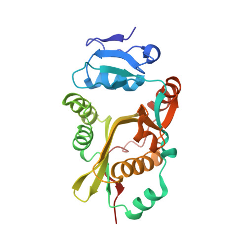
2Z3L

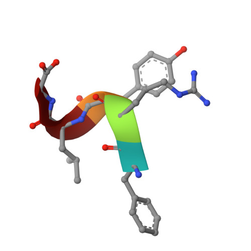
complex structure of LF-transferase and peptide A


Experimental Data Snapshot

  • Method: X-RAY DIFFRACTION
  • Resolution: 2.75 Å
  • R-Value Free: 0.265 
  • R-Value Work: 0.207 
  • R-Value Observed: 0.207 

Starting Model: experimental
View more details

wwPDB Validation   3D Report Full Report


This is version 1.2 of the entry. See complete history


Literature

Protein-based peptide-bond formation by aminoacyl-tRNA protein transferase

Watanabe, K.Toh, Y.Suto, K.Shimizu, Y.Oka, N.Wada, T.Tomita, K.

(2007) Nature 449: 867-871

  • DOI: https://doi.org/10.1038/nature06167
  • Primary Citation of Related Structures:  
    2Z3K, 2Z3L, 2Z3M, 2Z3N, 2Z3O, 2Z3P

  • PubMed Abstract: 

    Eubacterial leucyl/phenylalanyl-tRNA protein transferase (LF-transferase) catalyses peptide-bond formation by using Leu-tRNA(Leu) (or Phe-tRNA(Phe)) and an amino-terminal Arg (or Lys) of a protein, as donor and acceptor substrates, respectively. However, the catalytic mechanism of peptide-bond formation by LF-transferase remained obscure. Here we determine the structures of complexes of LF-transferase and phenylalanyl adenosine, with and without a short peptide bearing an N-terminal Arg. Combining the two separate structures into one structure as well as mutation studies reveal the mechanism for peptide-bond formation by LF-transferase. The electron relay from Asp 186 to Gln 188 helps Gln 188 to attract a proton from the alpha-amino group of the N-terminal Arg of the acceptor peptide. This generates the attacking nucleophile for the carbonyl carbon of the aminoacyl bond of the aminoacyl-tRNA, thus facilitating peptide-bond formation. The protein-based mechanism for peptide-bond formation by LF-transferase is similar to the reverse reaction of the acylation step observed in the peptide hydrolysis reaction by serine proteases.


  • Organizational Affiliation

    Institute of Biological Resources and Functions, National Institute of Advanced Industrial Sciences and Technology, 1-1-1, Higashi, Tsukuba-shi, Ibaraki 305-8566, Japan.


Macromolecules
Find similar proteins by:  (by identity cutoff)  |  3D Structure
Entity ID: 1
MoleculeChains Sequence LengthOrganismDetailsImage
Leucyl/phenylalanyl-tRNA-protein transferase
A, B
233Escherichia coliMutation(s): 0 
Gene Names: Aat
EC: 2.3.2.6
UniProt
Find proteins for P0A8P1 (Escherichia coli (strain K12))
Explore P0A8P1 
Go to UniProtKB:  P0A8P1
Entity Groups  
Sequence Clusters30% Identity50% Identity70% Identity90% Identity95% Identity100% Identity
UniProt GroupP0A8P1
Sequence Annotations
Expand
  • Reference Sequence

Find similar proteins by:  Sequence   |   3D Structure  

Entity ID: 2
MoleculeChains Sequence LengthOrganismDetailsImage
peptide (PHE)(ARG)(TYR)(LEU)(GLY)
C, D
5N/AMutation(s): 0 
Sequence Annotations
Expand
  • Reference Sequence
Experimental Data & Validation

Experimental Data

  • Method: X-RAY DIFFRACTION
  • Resolution: 2.75 Å
  • R-Value Free: 0.265 
  • R-Value Work: 0.207 
  • R-Value Observed: 0.207 
  • Space Group: P 21 21 2
Unit Cell:
Length ( Å )Angle ( ˚ )
a = 115.54α = 90
b = 129.998β = 90
c = 38.436γ = 90
Software Package:
Software NamePurpose
CNSrefinement
HKL-2000data scaling
CNSphasing

Structure Validation

View Full Validation Report


Entry History 

Deposition Data

Revision History  (Full details and data files)

  • Version 1.0: 2007-10-23
    Type: Initial release
  • Version 1.1: 2011-07-13
    Changes: Version format compliance
  • Version 1.2: 2023-11-01
    Changes: Data collection, Database references, Refinement description