


Crystal structure of VioE, a key player in the construction of the molecular skeleton of violacein
Hirano, S., Asamizu, S., Onaka, H., Shiro, Y., Nagano, S.To be published.
Experimental Data Snapshot
Starting Model: experimental
View more details
Entity ID: 1 | |||||
|---|---|---|---|---|---|
| Molecule | Chains | Sequence Length | Organism | Details | Image |
| Hypothetical protein VioE | 194 | Chromobacterium violaceum | Mutation(s): 0 | 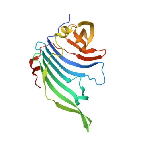 | |
UniProt | |||||
Entity Groups | |||||
| Sequence Clusters | 30% Identity50% Identity70% Identity90% Identity95% Identity100% Identity | ||||
| UniProt Group | Q7NSZ5 | ||||
Sequence AnnotationsExpand | |||||
Reference Sequence | |||||
| Ligands 1 Unique | |||||
|---|---|---|---|---|---|
| ID | Chains | Name / Formula / InChI Key | 2D Diagram | 3D Interactions | |
| PPY Download:Ideal Coordinates CCD File | G [auth A] H [auth A] I [auth B] J [auth B] K [auth C] | 3-PHENYLPYRUVIC ACID C9 H8 O3 BTNMPGBKDVTSJY-UHFFFAOYSA-N |  | ||
| Length ( Å ) | Angle ( ˚ ) |
|---|---|
| a = 83.415 | α = 90 |
| b = 91.03 | β = 90 |
| c = 158.999 | γ = 90 |
| Software Name | Purpose |
|---|---|
| REFMAC | refinement |
| HKL-2000 | data collection |
| HKL-2000 | data reduction |
| HKL-2000 | data scaling |
| MOLREP | phasing |


