2ZKN | pdb_00002zkn

X-ray structure of mutant galectin-1/lactose complex


Experimental Data Snapshot

  • Method: X-RAY DIFFRACTION
  • Resolution: 1.86 Å
  • R-Value Free: 
    0.244 (Depositor), 0.231 (DCC) 
  • R-Value Work: 
    0.215 (Depositor), 0.203 (DCC) 
  • R-Value Observed: 
    0.215 (Depositor) 

Starting Model: experimental
View more details

wwPDB Validation 3D Report Full Report

Validation slider image for 2ZKN

This is version 2.2 of the entry. See complete history

Literature

Functional and structural bases of a cysteine-less mutant as a long-lasting substitute for galectin-1

Nishi, N.Abe, A.Iwaki, J.Yoshida, H.Itoh, A.Shoji, H.Kamitori, S.Hirabayashi, J.Nakamura, T.

(2008) Glycobiology 18: 1065-1073

  • DOI: https://doi.org/10.1093/glycob/cwn089
  • Primary Citation Related Structures: 
    2ZKN

  • PubMed Abstract: 

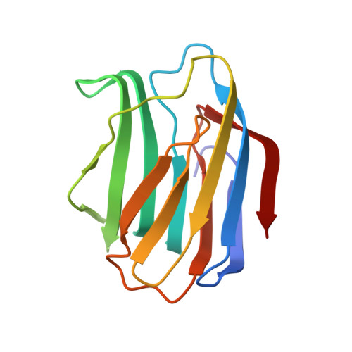
    Galectin-1 (Gal-1), a member of the beta-galactoside-binding animal lectin family, has a wide range of biological activities, which makes it an attractive target for medical applications. Unlike other galectins, Gal-1 is susceptible to oxidation at cysteine residues, which is troublesome for in vitro/vivo studies. To overcome this problem, we prepared a cysteine-less mutant of Gal-1 (CSGal-1) by substituting all cysteine residues with serine residues. In the case of wild-type Gal-1, the formation of covalent dimers/oligomers was evident after 10 days of storage in the absence of a reducing agent with a concomitant decrease in hemagglutination activity, while CSGal-1 did not form multimers and retained full hemagglutination activity after 400 days of storage. Frontal affinity chromatography showed that the sugar-binding specificity and affinity of Gal-1 for model glycans were barely affected by the mutagenesis. Gal-1 is known to induce cell signaling leading to an increase in the intracytoplasmic calcium concentration and to cell death. CSGal-1 is also capable of inducing calcium flux and growth inhibition in Jurkat cells, which are comparable to or more potent than those induced by Gal-1. The X-ray structure of the CSGal-1/lactose complex has been determined. The structure of CSGal-1 is almost identical to that of wild-type human Gal-1, showing that the amino acid substitutions do not affect the overall structure or carbohydrate-binding site structure of the protein. These results indicate that CSGal-1 can serve as a stable substitute for Gal-1.


  • Organizational Affiliation
    • Department of Endocrinology, Faculty of Medicine, Kagawa University, Kagawa, Japan. nnishi@med.kagawa-u.ac.jp

Macromolecule Content 

  • Total Structure Weight: 29.35 kDa 
  • Atom Count: 2,220 
  • Modeled Residue Count: 268 
  • Deposited Residue Count: 268 
  • Unique protein chains: 1

Macromolecules

Find similar proteins by:|  3D Structure
Entity ID: 1
MoleculeChains  Sequence LengthOrganismDetailsImage
Galectin-1
A, B
134Homo sapiensMutation(s): 6 
Gene Names: LGALS1
UniProt & NIH Common Fund Data Resources
Find proteins for P09382 (Homo sapiens)
Explore P09382 
Go to UniProtKB:  P09382
PHAROS:  P09382
GTEx:  ENSG00000100097 
Entity Groups
Sequence Clusters30% Identity50% Identity70% Identity90% Identity95% Identity100% Identity
UniProt GroupP09382
Sequence Annotations
Expand
Reference Sequence

Oligosaccharides

Help  
Entity ID: 2
MoleculeChains Length2D Diagram GlycosylationD Interactions
beta-D-galactopyranose-(1-4)-beta-D-glucopyranose
C
2N/A
Glycosylation Resources
GlyTouCan: G84224TW
GlyCosmos: G84224TW
GlyGen: G84224TW

Biologically Interesting Molecules (External Reference) 

1 Unique

Experimental Data & Validation

Experimental Data

  • Method: X-RAY DIFFRACTION
  • Resolution: 1.86 Å
  • R-Value Free:  0.244 (Depositor), 0.231 (DCC) 
  • R-Value Work:  0.215 (Depositor), 0.203 (DCC) 
  • R-Value Observed: 0.215 (Depositor) 
Space Group: P 21 21 21
Unit Cell:
Length ( Å )Angle ( ˚ )
a = 36.925α = 90
b = 87.431β = 90
c = 95.698γ = 90
Software Package:
Software NamePurpose
CNSrefinement
ADSCdata collection
HKL-2000data reduction
HKL-2000data scaling
CNSphasing

Structure Validation

View Full Validation Report


Entry History 

Deposition Data

Revision History  (Full details and data files)

  • Version 1.0: 2008-09-30
    Type: Initial release
  • Version 1.1: 2011-07-13
    Changes: Version format compliance
  • Version 2.0: 2020-07-29
    Type: Remediation
    Reason: Carbohydrate remediation
    Changes: Atomic model, Data collection, Derived calculations, Structure summary
  • Version 2.1: 2021-11-10
    Changes: Database references, Structure summary
  • Version 2.2: 2023-11-01
    Changes: Data collection, Refinement description