2ZLD | pdb_00002zld

Structure of OmpF co-crystallized with T83


Experimental Data Snapshot

  • Method: X-RAY DIFFRACTION
  • Resolution: 3.00 Å
  • R-Value Free: 
    0.294 (Depositor) 
  • R-Value Work: 
    0.266 (Depositor) 
  • R-Value Observed: 
    0.267 (Depositor) 

Starting Model: experimental
View more details

wwPDB Validation 3D Report Full Report

Validation slider image for 2ZLD

This is version 1.2 of the entry. See complete history

Literature

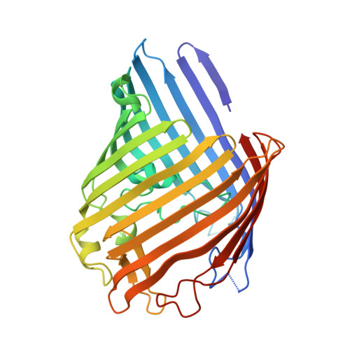
Crystal structures of the OmpF porin: function in a colicin translocon.

Yamashita, E.Zhalnina, M.V.Zakharov, S.D.Sharma, O.Cramer, W.A.

(2008) EMBO J 27: 2171-2180

  • DOI: https://doi.org/10.1038/emboj.2008.137
  • Primary Citation Related Structures: 
    2ZFG, 2ZLD

  • PubMed Abstract: 

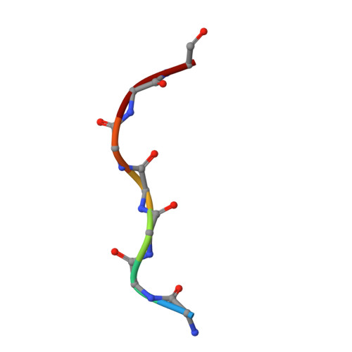
    The OmpF porin in the Escherichia coli outer membrane (OM) is required for the cytotoxic action of group A colicins, which are proposed to insert their translocation and active domains through OmpF pores. A crystal structure was sought of OmpF with an inserted colicin segment. A 1.6 A OmpF structure, obtained from crystals formed in 1 M Mg2+, has one Mg2+ bound in the selectivity filter between Asp113 and Glu117 of loop 3. Co-crystallization of OmpF with the unfolded 83 residue glycine-rich N-terminal segment of colicin E3 (T83) that occludes OmpF ion channels yielded a 3.0 A structure with inserted T83, which was obtained without Mg2+ as was T83 binding to OmpF. The incremental electron density could be modelled as an extended poly-glycine peptide of at least seven residues. It overlapped the Mg2+ binding site obtained without T83, explaining the absence of peptide binding in the presence of Mg2+. Involvement of OmpF in colicin passage through the OM was further documented by immuno-extraction of an OM complex, the colicin translocon, consisting of colicin E3, BtuB and OmpF.


  • Organizational Affiliation
    • Department of Biological Sciences, Purdue University, West Lafayette, IN 47907-1392, USA.

Macromolecule Content 

  • Total Structure Weight: 75.46 kDa 
  • Atom Count: 5,294 
  • Modeled Residue Count: 692 
  • Deposited Residue Count: 694 
  • Unique protein chains: 2

Macromolecules

Find similar proteins by:|  3D Structure
Entity ID: 1
MoleculeChains  Sequence LengthOrganismDetailsImage
Outer membrane protein F
A, B
340Escherichia coliMutation(s): 0 
Gene Names: ompFcmlBcoacrytolFb0929JW0912
Membrane Entity: Yes 
UniProt
Find proteins for P02931 (Escherichia coli (strain K12))
Explore P02931 
Go to UniProtKB:  P02931
Entity Groups
Sequence Clusters30% Identity50% Identity70% Identity90% Identity95% Identity100% Identity
UniProt GroupP02931
Sequence Annotations
Expand
Reference Sequence
Find similar proteins by:  Sequence   |   3D Structure  
Entity ID: 2
MoleculeChains  Sequence LengthOrganismDetailsImage
Colicin-E3
C, D
7Escherichia coliMutation(s): 0 
EC: 3.1

Experimental Data & Validation

Experimental Data

  • Method: X-RAY DIFFRACTION
  • Resolution: 3.00 Å
  • R-Value Free:  0.294 (Depositor) 
  • R-Value Work:  0.266 (Depositor) 
  • R-Value Observed: 0.267 (Depositor) 
Space Group: P 63
Unit Cell:
Length ( Å )Angle ( ˚ )
a = 116.867α = 90
b = 116.867β = 90
c = 114.331γ = 120
Software Package:
Software NamePurpose
REFMACrefinement
HKL-3000data reduction
SCALEPACKdata scaling
MOLREPphasing

Structure Validation

View Full Validation Report


Entry History 

Deposition Data

Revision History  (Full details and data files)

  • Version 1.0: 2008-07-29
    Type: Initial release
  • Version 1.1: 2011-07-13
    Changes: Version format compliance
  • Version 1.2: 2023-11-01
    Changes: Data collection, Database references, Refinement description