2ZOX | pdb_00002zox

Crystal Structure of the Covalent Intermediate of Human Cytosolic beta-Glucosidase


Experimental Data Snapshot

  • Method: X-RAY DIFFRACTION
  • Resolution: 1.90 Å
  • R-Value Free: 
    0.226 (Depositor), 0.205 (DCC) 
  • R-Value Work: 
    0.191 (Depositor) 
  • R-Value Observed: 
    0.192 (Depositor) 

Starting Model: experimental
View more details

wwPDB Validation 3D Report Full Report

Validation slider image for 2ZOX

Ligand Structure Quality Assessment 


This is version 1.5 of the entry. See complete history

Literature

Crystal structure of the covalent intermediate of human cytosolic beta-glucosidase

Noguchi, J.Hayashi, Y.Baba, Y.Okino, N.Kimura, M.Ito, M.Kakuta, Y.

(2008) Biochem Biophys Res Commun 374: 549-552

  • DOI: https://doi.org/10.1016/j.bbrc.2008.07.089
  • Primary Citation Related Structures: 
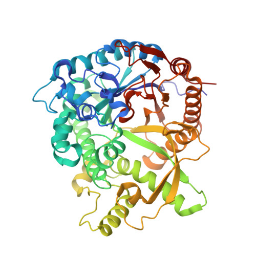
    2ZOX

  • PubMed Abstract: 

    Human cytosolic beta-glucosidase, also known as klotho-related protein (KLrP, GBA3), is an enzyme that hydrolyzes various beta-D-glucosides, including glucosylceramide. We recently reported the crystal structure of KLrP in complex with glucose [Y. Hayashi, N. Okino, Y. Kakuta, T. Shikanai, M. Tani, H. Narimatsu, M. Ito, Klotho-related protein is a novel cytosolic neutral beta-glycosylceramidase, J. Biol. Chem. 282 (2007) 30889-30900]. Here, we report the crystal structure of a covalent intermediate of the KLrP mutant E165Q, in which glucose was covalently bound to a nucleophile, Glu(373). The structure confirms the double displacement mechanism of the retaining beta-glucosidase. In addition, the structure suggests that a water molecule could be involved in the stabilization of transition states through a sugar, 2-hydroxyl.


  • Organizational Affiliation
    • Laboratory of Structural Biology, Graduate School of Systems Life Sciences, Kyushu University, Fukuoka 812-8581, Japan.

Macromolecule Content 

  • Total Structure Weight: 55.26 kDa 
  • Atom Count: 4,092 
  • Modeled Residue Count: 466 
  • Deposited Residue Count: 469 
  • Unique protein chains: 1

Macromolecules

Find similar proteins by:|  3D Structure
Entity ID: 1
MoleculeChains  Sequence LengthOrganismDetailsImage
Cytosolic beta-glucosidase469Homo sapiensMutation(s): 1 
Gene Names: GBA3CBGCBGL1
EC: 3.2.1.21 (PDB Primary Data), 3.2.1.46 (UniProt), 3.2.1.45 (UniProt)
UniProt & NIH Common Fund Data Resources
Find proteins for Q9H227 (Homo sapiens)
Explore Q9H227 
Go to UniProtKB:  Q9H227
PHAROS:  Q9H227
Entity Groups
Sequence Clusters30% Identity50% Identity70% Identity90% Identity95% Identity100% Identity
UniProt GroupQ9H227
Sequence Annotations
Expand
Reference Sequence

Small Molecules

Ligands 7 Unique
IDChains Name / Formula / InChI Key2D Diagram3D Interactions
PNG

Query on PNG


Download:Ideal Coordinates CCD File
C [auth A]4-nitrophenyl alpha-D-glucopyranoside
C12 H15 N O8
IFBHRQDFSNCLOZ-ZIQFBCGOSA-N
OLA

Query on OLA


Download:Ideal Coordinates CCD File
H [auth A]OLEIC ACID
C18 H34 O2
ZQPPMHVWECSIRJ-KTKRTIGZSA-N
PLM

Query on PLM


Download:Ideal Coordinates CCD File
G [auth A],
I [auth A]
PALMITIC ACID
C16 H32 O2
IPCSVZSSVZVIGE-UHFFFAOYSA-N
GLC

Query on GLC


Download:Ideal Coordinates CCD File
B [auth A]alpha-D-glucopyranose
C6 H12 O6
WQZGKKKJIJFFOK-DVKNGEFBSA-N
PO4

Query on PO4


Download:Ideal Coordinates CCD File
F [auth A]PHOSPHATE ION
O4 P
NBIIXXVUZAFLBC-UHFFFAOYSA-K
GOL

Query on GOL


Download:Ideal Coordinates CCD File
J [auth A]GLYCEROL
C3 H8 O3
PEDCQBHIVMGVHV-UHFFFAOYSA-N
MG

Query on MG


Download:Ideal Coordinates CCD File
D [auth A],
E [auth A]
MAGNESIUM ION
Mg
JLVVSXFLKOJNIY-UHFFFAOYSA-N

Experimental Data & Validation

Experimental Data

  • Method: X-RAY DIFFRACTION
  • Resolution: 1.90 Å
  • R-Value Free:  0.226 (Depositor), 0.205 (DCC) 
  • R-Value Work:  0.191 (Depositor) 
  • R-Value Observed: 0.192 (Depositor) 
Space Group: P 21 21 21
Unit Cell:
Length ( Å )Angle ( ˚ )
a = 66.12α = 90
b = 82.529β = 90
c = 91.673γ = 90
Software Package:
Software NamePurpose
REFMACrefinement
HKL-2000data reduction
HKL-2000data scaling
MOLREPphasing

Structure Validation

View Full Validation Report


Ligand Structure Quality Assessment 


Entry History 

Deposition Data

Revision History  (Full details and data files)

  • Version 1.0: 2008-09-30
    Type: Initial release
  • Version 1.1: 2011-07-13
    Changes: Non-polymer description, Version format compliance
  • Version 1.2: 2020-07-29
    Type: Remediation
    Reason: Carbohydrate remediation
    Changes: Advisory, Data collection, Derived calculations, Structure summary
  • Version 1.3: 2021-11-10
    Changes: Database references, Structure summary
  • Version 1.4: 2023-11-01
    Changes: Data collection, Refinement description
  • Version 1.5: 2024-10-30
    Changes: Structure summary