2ZVJ | pdb_00002zvj

Crystal structures of rat Catechol-O-Methyltransferase complexed with coumarine-based inhibitor


Experimental Data Snapshot

  • Method: X-RAY DIFFRACTION
  • Resolution: 2.30 Å
  • R-Value Free: 
    0.286 (Depositor), 0.285 (DCC) 
  • R-Value Work: 
    0.211 (Depositor), 0.213 (DCC) 
  • R-Value Observed: 
    0.219 (Depositor) 

Starting Model: experimental
View more details

wwPDB Validation 3D Report Full Report

Validation slider image for 2ZVJ

Ligand Structure Quality Assessment 


This is version 1.2 of the entry. See complete history

Literature

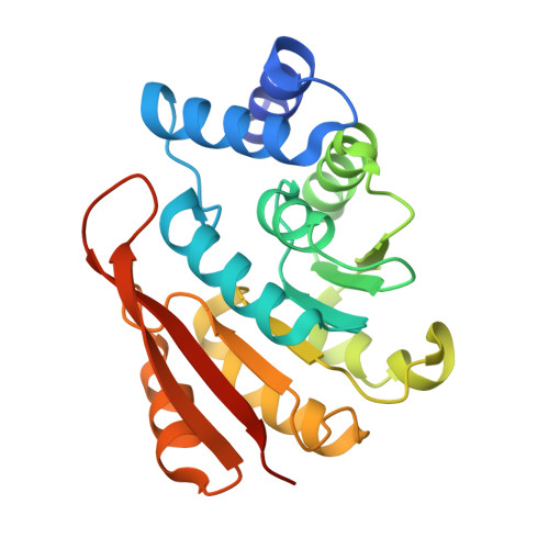
Crystal structures of rat catechol-O-methyltransferase complexed with coumarine-based inhibitor

Tsuji, E.Okazaki, K.Takeda, K.

(2009) Biochem Biophys Res Commun 378: 494-497

  • DOI: https://doi.org/10.1016/j.bbrc.2008.11.085
  • Primary Citation Related Structures: 
    2ZVJ

  • PubMed Abstract: 

    In human, catechol-O-methyltransferase (COMT: E.C. 2.1.1.6) is responsible for metabolism of catechol neurotransmitter and xenobiotics. The main clinical interest in COMT results from the possibility of using COMT inhibitors as adjuncts in the therapy of Parkinson's disease (PD) with l-DOPA. COMT is therefore a target for inhibitor development aiming at PD treatment and has been submitted to extensive structure-based drug design. Recently reported inhibitors have nitrocatechol structure that may inhibit oxidative phosphorylation and uncouple mitochondrial energy production. This work reports the first crystallographic study of Rat COMT complexed with non-nitrocatechol inhibitor. Analysis of the structural differences among the previously reported inhibitor complexes, coumarine-based inhibitor (4-phenyl-7, 8-dihydroxycoumarine: 4PCM) bound structure provides the explanation for inhibitor binding and can be used for future inhibitor design.


  • Organizational Affiliation
    • Molecular Design Research, R&D Kissei Pharmaceutical Co., Ltd. 4365-1 Kashiwabara, Hotaka, Azumino-city, Nagano 399-8304, Japan.

Macromolecule Content 

  • Total Structure Weight: 25.59 kDa 
  • Atom Count: 1,792 
  • Modeled Residue Count: 212 
  • Deposited Residue Count: 223 
  • Unique protein chains: 1

Macromolecules

Find similar proteins by:|  3D Structure
Entity ID: 1
MoleculeChains  Sequence LengthOrganismDetailsImage
Catechol O-methyltransferase223Rattus norvegicusMutation(s): 0 
EC: 2.1.1.6
UniProt
Find proteins for P22734 (Rattus norvegicus)
Explore P22734 
Go to UniProtKB:  P22734
Entity Groups
Sequence Clusters30% Identity50% Identity70% Identity90% Identity95% Identity100% Identity
UniProt GroupP22734
Sequence Annotations
Expand
Reference Sequence

Experimental Data & Validation

Experimental Data

  • Method: X-RAY DIFFRACTION
  • Resolution: 2.30 Å
  • R-Value Free:  0.286 (Depositor), 0.285 (DCC) 
  • R-Value Work:  0.211 (Depositor), 0.213 (DCC) 
  • R-Value Observed: 0.219 (Depositor) 
Space Group: P 32 2 1
Unit Cell:
Length ( Å )Angle ( ˚ )
a = 50.448α = 90
b = 50.448β = 90
c = 167.626γ = 120
Software Package:
Software NamePurpose
REFMACrefinement
BBSdata collection
MOSFLMdata reduction
SCALAdata scaling
AMoREphasing

Structure Validation

View Full Validation Report


Ligand Structure Quality Assessment 


Entry History 

Deposition Data

  • Released Date: 2009-01-06 
  • Deposition Author(s): Tsuji, E.

Revision History  (Full details and data files)

  • Version 1.0: 2009-01-06
    Type: Initial release
  • Version 1.1: 2011-07-13
    Changes: Version format compliance
  • Version 1.2: 2023-11-01
    Changes: Data collection, Database references, Derived calculations, Refinement description