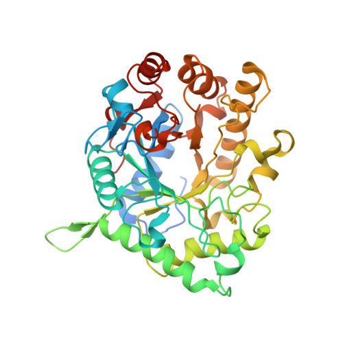
The Cellvibrio Japonicus Mannanase Cjman26C Displays a Unique Exo-Mode of Action that is Conferred by Subtle Changes to the Distal Region of the Active Site.
Cartmell, A., Topakas, E., Ducros, V.M.-A., Suits, M.D.L., Davies, G.J., Gilbert, H.J.(2008) J Biol Chem 283: 34403
- PubMed: 18799462
- DOI: https://doi.org/10.1074/jbc.M804053200
- Primary Citation of Related Structures:
2VX4, 2VX5, 2VX6, 2VX7 - PubMed Abstract:
The microbial degradation of the plant cell wall is a pivotal biological process that is of increasing industrial significance. One of the major plant structural polysaccharides is mannan, a beta-1,4-linked d-mannose polymer, which is hydrolyzed by endo- and exo-acting mannanases. The mechanisms by which the exo-acting enzymes target the chain ends of mannan and how galactose decorations influence activity are poorly understood. Here we report the crystal structure and biochemical properties of CjMan26C, a Cellvibrio japonicus GH26 mannanase. The exo-acting enzyme releases the disaccharide mannobiose from the nonreducing end of mannan and mannooligosaccharides, harnessing four mannose-binding subsites extending from -2 to +2. The structure of CjMan26C is very similar to that of the endo-acting C. japonicus mannanase CjMan26A. The exo-activity displayed by CjMan26C, however, reflects a subtle change in surface topography in which a four-residue extension of surface loop creates a steric block at the distal glycone -2 subsite. endo-Activity can be introduced into enzyme variants through truncation of an aspartate side chain, a component of a surface loop, or by removing both the aspartate and its flanking residues. The structure of catalytically competent CjMan26C, in complex with a decorated manno-oligosaccharide, reveals a predominantly unhydrolyzed substrate in an approximate (1)S(5) conformation. The complex structure helps to explain how the substrate "side chain" decorations greatly reduce the activity of the enzyme; the galactose side chain at the -1 subsite makes polar interactions with the aglycone mannose, possibly leading to suboptimal binding and impaired leaving group departure. This report reveals how subtle differences in the loops surrounding the active site of a glycoside hydrolase can lead to a change in the mode of action of the enzyme.
Organizational Affiliation:
Institute for Cell and Molecular Biosciences, The Medical School, Newcastle University, Framlington Place, Newcastle upon Tyne NE2 4HH, United Kingdom.


