37PY | pdb_000037py

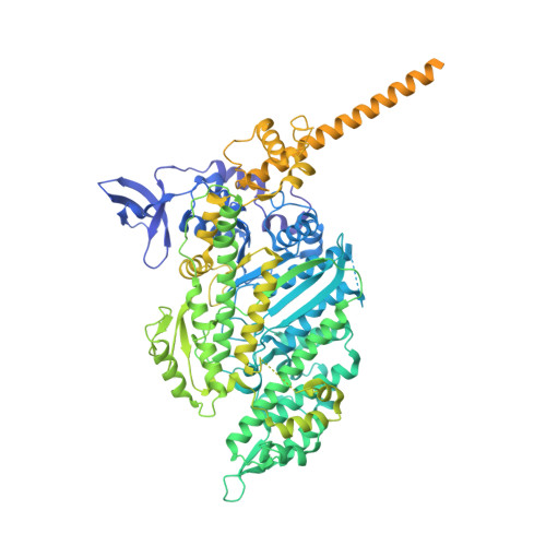
Human beta-cardiac myosin S1 motor domain with the essential light chain in the pre-powerstroke state


Experimental Data Snapshot

  • Method: ELECTRON MICROSCOPY
  • Resolution: 3.20 Å
  • Aggregation State: PARTICLE 
  • Reconstruction Method: SINGLE PARTICLE 

Starting Model: experimental
View more details

wwPDB Validation 3D Report Full Report

Validation slider image for 37PY

This is version 1.0 of the entry. See complete history. 

Literature

The dilated cardiomyopathy E525K mutation stabilizes the cardiac myosin interacting-heads motif while activating the isolated motor domain.

Gautam, R., Somavarapu, A.K., Robeson, K.Z., Childers, M.C., Regnier, M., Ge, J., Yengo, C.M., Craig, R., Padron, R.

(2026) bioRxiv 

  • DOI: https://doi.org/10.64898/2026.10.02.756299
  • Primary Citation Related Structures: 
    37OA, 37PY, 37QC

  • PubMed Abstract: 

    Mutations in β-cardiac myosin are a common cause of inherited cardiomyopathies. The dilated cardiomyopathy E525K mutation alters both thick filament regulation and motor activity, yet the structural basis for these effects has remained unclear. Here, we combined cryo-EM and MD simulations to determine how E525K affects the conformational landscape of cardiac myosin in both its autoinhibited interacting-heads motif (IHM) and isolated myosin head states. E525K-mutated myosin exclusively adopted a single S2 conformation in IHM, in contrast to the conformational heterogeneity observed in wild-type myosin. Structural and electrostatic analyses revealed that the mutation increased positive charge density at the blocked-head (BH) - S2 (subfragment-2) interface, strengthening interactions with negatively charged residues within S2. 3D variability analysis and MD simulations of the E525K IHM demonstrated reduced S2 mobility, consistent with strengthened BH - S2 interactions, and decreased conformational flexibility, providing a structural mechanism for stabilization of the IHM state. To investigate the isolated myosin head, we determined cryo-EM structures of wild-type and E525K subfragment-1 (S1). The mutation induced local conformational rearrangements in the activation-loop and Loop 3 region, the SH3-like domain, and the essential light chain of S1, revealing structural changes that support the enhanced motor activity previously reported for isolated E525K myosin motors. These findings show that E525K stabilizes the autoinhibited IHM through electrostatic stabilization of the BH - S2 interface while inducing structural changes consistent with activation of the isolated motor domain, providing structural insights into how a single DCM mutation differentially regulates cardiac myosin S1 and IHM structure and function.


  • Organizational Affiliation: 
    • Division of Cell Biology and Imaging, Department of Radiology, University of Massachusetts Chan Medical School; Worcester, MA, USA.

Macromolecule Content 

  • Total Structure Weight: 147.65 kDa 
  • Atom Count: 7,363 
  • Modeled Residue Count: 914 
  • Deposited Residue Count: 1,297 
  • Unique protein chains: 2

Macromolecules

Find similar proteins by:|  3D Structure
Entity ID: 1
MoleculeChains  Sequence LengthOrganismDetailsImage
Myosin-7,Green fluorescent protein1,109Homo sapiens, Aequorea victoria
This entity is chimeric
Mutation(s): 0 
Gene Names: MYH7, MYHCB, GFP
UniProt & NIH Common Fund Data Resources
Find proteins for P12883 (Homo sapiens)
Explore P12883 
Go to UniProtKB:  P12883
PHAROS:  P12883
GTEx:  ENSG00000092054 
Find proteins for P42212 (Aequorea victoria)
Explore P42212 
Go to UniProtKB:  P42212
Entity Groups
Sequence Clusters30% Identity50% Identity70% Identity90% Identity95% Identity100% Identity
UniProt GroupsP12883P42212
Sequence Annotations
Expand
Reference Sequence
Find similar proteins by:|  3D Structure
Entity ID: 2
MoleculeChains  Sequence LengthOrganismDetailsImage
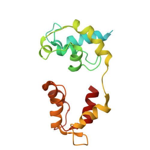
Myosin light chain 1/3, skeletal muscle isoform188Mus musculusMutation(s): 0 
UniProt
Find proteins for P05977 (Mus musculus)
Explore P05977 
Go to UniProtKB:  P05977
Entity Groups
Sequence Clusters30% Identity50% Identity70% Identity90% Identity95% Identity100% Identity
UniProt GroupP05977
Sequence Annotations
Expand
Reference Sequence

Experimental Data & Validation

Experimental Data

  • Method: ELECTRON MICROSCOPY
  • Resolution: 3.20 Å
  • Aggregation State: PARTICLE 
  • Reconstruction Method: SINGLE PARTICLE 
EM Software:
TaskSoftware PackageVersion
RECONSTRUCTIONcryoSPARC
MODEL REFINEMENTPHENIX1.20.1_4487

Structure Validation

View Full Validation Report


Entry History 

& Funding Information

Deposition Data


Funding OrganizationLocationGrant Number
National Institutes of Health/National Heart, Lung, and Blood Institute (NIH/NHLBI)United StatesHL164560
National Institutes of Health/National Institute of Arthritis and Musculoskeletal and Skin Diseases (NIH/NIAMS)United StatesAR081941
American Heart AssociationUnited States26CDA1600784

Revision History  (Full details and data files)

  • Version 1.0: 2026-11-04
    Type: Initial release